. 2009 Dec 29:9:465.
doi: 10.1186/1471-2407-9-465.

A reproducible brain tumour model established from human glioblastoma biopsies

Affiliations

A reproducible brain tumour model established from human glioblastoma biopsies

Jian Wang et al. BMC Cancer. .

Abstract

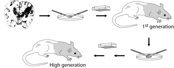
Background: Establishing clinically relevant animal models of glioblastoma multiforme (GBM) remains a challenge, and many commonly used cell line-based models do not recapitulate the invasive growth patterns of patient GBMs. Previously, we have reported the formation of highly invasive tumour xenografts in nude rats from human GBMs. However, implementing tumour models based on primary tissue requires that these models can be sufficiently standardised with consistently high take rates.

Methods: In this work, we collected data on growth kinetics from a material of 29 biopsies xenografted in nude rats, and characterised this model with an emphasis on neuropathological and radiological features.

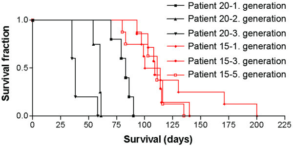
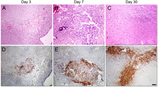
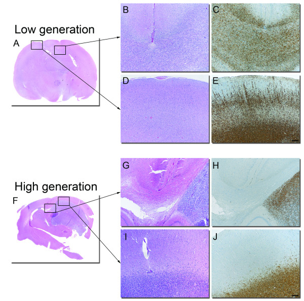
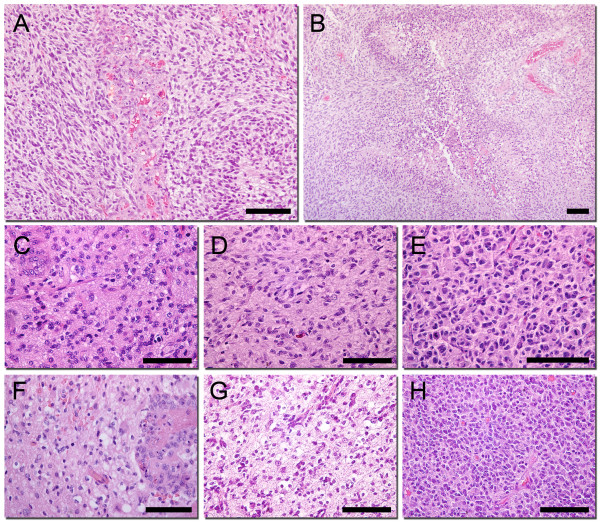
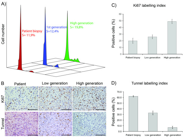
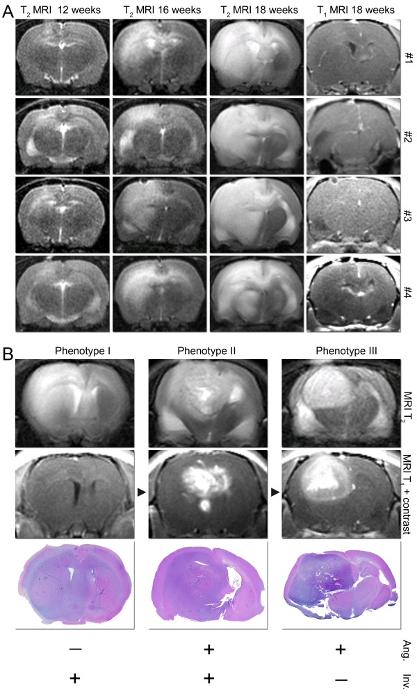
Results: The tumour take rate for xenografted GBM biopsies were 96% and remained close to 100% at subsequent passages in vivo, whereas only one of four lower grade tumours engrafted. Average time from transplantation to the onset of symptoms was 125 days +/- 11.5 SEM. Histologically, the primary xenografts recapitulated the invasive features of the parent tumours while endothelial cell proliferations and necrosis were mostly absent. After 4-5 in vivo passages, the tumours became more vascular with necrotic areas, but also appeared more circumscribed. MRI typically revealed changes related to tumour growth, several months prior to the onset of symptoms.

Conclusions: In vivo passaging of patient GBM biopsies produced tumours representative of the patient tumours, with high take rates and a reproducible disease course. The model provides combinations of angiogenic and invasive phenotypes and represents a good alternative to in vitro propagated cell lines for dissecting mechanisms of brain tumour progression.


Figures

Figure 1
Figure 1
Experimental design. Biopsy tissues from human gliomas are minced into small fragments that are subsequently cultured in medium. The tumour fragments remodel and form small tumour spheroids, usually within 1-2 weeks. These spheroids are stereotactically implanted in the nude rat brain, and form xenograft tumours (1st or low generation) that are harvested and processed similarly to the biopsy tissue. The resulting spheroids are passaged onto a new generation of rats and the previous steps can be repeated to passage the tumour for several generations.
Figure 2
Figure 2
Survival as a function of tumour passaging in vivo. Shown are survival data for nude rats xenografted with GBM-biopsy tissue that were passaged in vivo for several generations. The patient number refers to cases listed in Table 1. For rats xenografted with tumour tissue from patient 20, survival was significantly shorter with passaging for 3 generations (p = 0.0029). For rats xenografted with tissue from patient 15, survival also decreased with passaging, although this was not significant (p = 0.69).
Figure 3
Figure 3
Xenograft tumour histology in initial stages of tumour growth. Growth of 1st generation tumours 3 (A), 7 (B) and 10 days (C) following implantation of patient biopsy spheroids, H/E staining. Shown are the implantation site (A, B) and contralateral hemisphere (C). Corresponding sections were also stained with a human-specific antibody against nestin (D, E, F). Scale bars = 100 μm
Figure 4
Figure 4
Xenograft tumour histology in early and late generations. Growth of 1st (A, B, C, D, E) and 10th generation (F, G, H, I, J) tumours in different areas of the nude rat brain. Corpus Callosum; H/E staining (B, G) and nestin staining (C, H), Cortex; H&E (D, I) and nestin (E, J). Scale bars = 250 μm.
Figure 5
Figure 5
Comparison of tumour cell morphology between patient and xenograft tumours. (A) Glomeruloid microvascular proliferations in a high generation tumour, H&E staining. (B) Palisading necrosis in a high generation tumour, H&E staining. Patient biopsies (C, F) with corresponding low (D, G) and high generation xenografts (E, H), H&E staining. Scale bars = 100 μm
Figure 6
Figure 6
Higher growth rates after passaging are mediated by increased tumour cell proliferation and reduced tumour cell death. S-phase fraction in a patient biopsy and the resulting xenograft tumours at various stages of passaging (A). Ki67 and Tunnel staining of corresponding tumour biopsies (B), with quantification of their labelling indexes (C) and (D), respectively. Scale bars = 100 μm.
Figure 7
Figure 7
MRI monitoring shows that brain tumour growth can be standardised and that the phenotypes are modulated with passaging in vivo. Coronary MRI scans showing tumour growth in four rats grafted with tissue from the same patient biopsy. Shown are T2 sequences at 3 different time points as indicated, while the right column present T1 images after administering contrast agent (A). MRI scans demonstrating different brain tumour phenotypes (B), displaying angiogenic and invasive growth as indicated (bottom). Corresponding H/E stained tumour sections are shown below the MRI panels. Extended passaging provide 3 distinct phenotypes that represent different combinations of invasive and angiogenic growth patterns.

References

    1. Ponten J, Macintyre EH. Long term culture of normal and neoplastic human glia. Acta Pathol Microbiol Scand. 1968;74(4):465–486. - PubMed
    1. Schold SC Jr, Friedman HS. Human brain tumor xenografts. Prog Exp Tumor Res. 1984;28:18–31. - PubMed
    1. Ross DT, Scherf U, Eisen MB, Perou CM, Rees C, Spellman P, Iyer V, Jeffrey SS, Rijn M Van de, Waltham M. Systematic variation in gene expression patterns in human cancer cell lines. Nat Genet. 2000;24(3):227–235. doi: 10.1038/73432. - DOI - PubMed
    1. Mahesparan R, Read TA, Lund-Johansen M, Skaftnesmo KO, Bjerkvig R, Engebraaten O. Expression of extracellular matrix components in a highly infiltrative in vivo glioma model. Acta Neuropathol. 2003;105(1):49–57. - PubMed
    1. Read TA, Farhadi M, Bjerkvig R, Olsen BR, Rokstad AM, Huszthy PC, Vajkoczy P. Intravital microscopy reveals novel antivascular and antitumor effects of endostatin delivered locally by alginate-encapsulated cells. Cancer Res. 2001;61(18):6830–6837. - PubMed

Publication types