Review
. 2009 Dec 29:8:69.
doi: 10.1186/1475-2859-8-69.

Tuning microbial hosts for membrane protein production

Affiliations
Review

Tuning microbial hosts for membrane protein production

Maria Freigassner et al. Microb Cell Fact. .

Abstract

The last four years have brought exciting progress in membrane protein research. Finally those many efforts that have been put into expression of eukaryotic membrane proteins are coming to fruition and enable to solve an ever-growing number of high resolution structures. In the past, many skilful optimization steps were required to achieve sufficient expression of functional membrane proteins. Optimization was performed individually for every membrane protein, but provided insight about commonly encountered bottlenecks and, more importantly, general guidelines how to alleviate cellular limitations during microbial membrane protein expression. Lately, system-wide analyses are emerging as powerful means to decipher cellular bottlenecks during heterologous protein production and their use in microbial membrane protein expression has grown in popularity during the past months.This review covers the most prominent solutions and pitfalls in expression of eukaryotic membrane proteins using microbial hosts (prokaryotes, yeasts), highlights skilful applications of our basic understanding to improve membrane protein production. Omics technologies provide new concepts to engineer microbial hosts for membrane protein production.


Figures
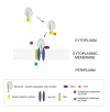
Figure 1
Figure 1
Membrane protein biogenesis in prokaryotes. In prokaryotes, most membrane proteins are targeted to and inserted into the cytoplasmic membrane by the SRP pathway, which involves interaction of the growing polypeptide chain with the signal recognition particle (SRP) and its receptor FtsY, binding of the nascent polypeptide chain-ribosome-complex to the SecYEG/YidC pore, translocation of cytoplasmic and periplasmic loops across the cytoplasmic membrane and their folding by SecA and various chaperones and insertion of hydrophobic segments into the membrane. The autonomous, YidC and Tat pathways, that are used by small proteins and membrane-associated periplasmic proteins, respectively, are mentioned here for sake of completeness.
Figure 2
Figure 2
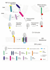
Membrane protein biogenesis in eukaryotes. In eukaryotic cells, membrane protein biogenesis occurs in a cotranslational way. Proteins residing in membranes of ER and Golgi apparatus or in plasma membrane use the secretory pathway. Like in prokaryotes, SRP recognizes polypeptides protruding from the ribosome complex, thereby transiently attenuating translation. As soon as the SRP-ribosome complex interacts with the SRP receptor and docks to the Sec61 translocon pore, translation resumes, BiP relocates, thereby opening the lumenal gate and the membrane protein enters the membrane by lateral diffusion through the Sec61 pore. Peroxisomal membrane proteins either use the Pex19/Pex3-mediated way (class I proteins, shown here) or are thought to reach the peroxisomal membrane via the ER (class II) [208]. Mitochondrial membrane proteins pass through the outer mitochondrial membrane (OMM) via the TOM complex (translocase of the outer membrane). While β-barrel proteins of the OMM are transported to the SAM and MDM complexes (sorting and assembly machinery), the TIM22 and TIM23 complexes (translocase of the inner mitochondrial membrane) are used to target proteins to the inner mitochondrial membrane [209].

Similar articles

Cited by

References

    1. Warne T, Serrano-Vega MJ, Baker JG, Moukhametzianov R, Edwards PC, Henderson R, Leslie AG, Tate CG, Schertler GF. Structure of a beta1-adrenergic G-protein-coupled receptor. Nature. 2008;454:486–491. doi: 10.1038/nature07101. - DOI - PMC - PubMed
    1. Rasmussen SG, Choi HJ, Rosenbaum DM, Kobilka TS, Thian FS, Edwards PC, Burghammer M, Ratnala VR, Sanishvili R, Fischetti RF, Schertler GF, Weis WI, Kobilka BK. Crystal structure of the human beta2 adrenergic G-protein-coupled receptor. Nature. 2007;450:383–387. doi: 10.1038/nature06325. - DOI - PubMed
    1. Cherezov V, Rosenbaum DM, Hanson MA, Rasmussen SG, Thian FS, Kobilka TS, Choi HJ, Kuhn P, Weis WI, Kobilka BK, Stevens RC. High-resolution crystal structure of an engineered human beta2-adrenergic G protein-coupled receptor. Science. 2007;318:1258–1265. doi: 10.1126/science.1150577. - DOI - PMC - PubMed
    1. Hanson MA, Cherezov V, Griffith MT, Roth CB, Jaakola VP, Chien EY, Velasquez J, Kuhn P, Stevens RC. A specific cholesterol binding site is established by the 2.8 A structure of the human beta2-adrenergic receptor. Structure. 2008;16:897–905. doi: 10.1016/j.str.2008.05.001. - DOI - PMC - PubMed
    1. Jaakola VP, Griffith MT, Hanson MA, Cherezov V, Chien EY, Lane JR, Ijzerman AP, Stevens RC. The 2.6 angstrom crystal structure of a human A2A adenosine receptor bound to an antagonist. Science. 2008;322:1211–1217. doi: 10.1126/science.1164772. - DOI - PMC - PubMed

Publication types

LinkOut - more resources