

Hippocampal subdivision and amygdalar volumes in patients in an at-risk mental state for schizophrenia
- PMID: 20040244
- PMCID: PMC2799502
- DOI: 10.1503/jpn.090013
Hippocampal subdivision and amygdalar volumes in patients in an at-risk mental state for schizophrenia
Abstract
Background: Accumulating evidence from postmortem and magnetic resonance imaging (MRI) studies suggests that abnormalities of medial temporal lobe structures are critically involved in the pathogenesis of schizophrenia. It is still unclear, however, whether certain abnormalities are already present in individuals at ultra high-risk (UHR) for transition into psychosis. Recent studies involving patients at UHR showed contradictory results for hippocampal volume, and only 1 study reported that amygdalar volume was unchanged between healthy patients and those at UHR. Furthermore, no subregions of the hippocampus have been investigated in people at UHR.
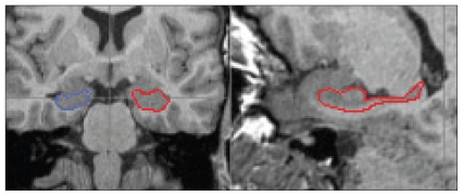
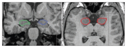
Methods: We recruited 29 UHR patients, 23 first-episode patients and 29 age- and sex-matched healthy controls. We measured hippocampal and amygdalar volumes from MRI scans by use of BRAINS2 to manually trace the regions of interest. The hippocampi were divided in 2 regions: head and corpus/tail.
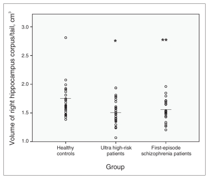
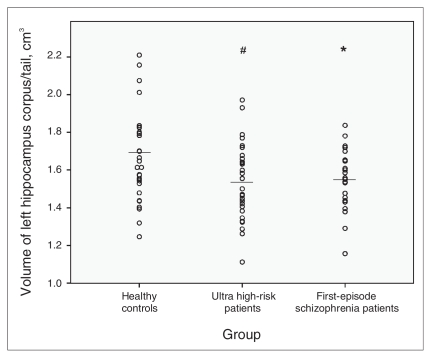
Results: Patients at UHR had significantly smaller volumes of the hippocampus corpus and tail bilaterally, but not of the head, compared with healthy controls. Group differences for the right hippocampus corpus and tail volume remained significant after we controlled for whole brain volume and other covariates. We found that UHR patients who later developed psychosis had smaller right hippocampus corpus and tail volumes than did those who did not develop psychosis. First-episode patients had significantly smaller left amygdalar volumes than did healthy individuals or those at UHR.
Limitations: Our study had a small sample size, and we were unable to control for the effects of medication.
Conclusion: Our findings suggest that parts of the hippocampal-amygdalar complex are involved in the pathogenesis of schizophrenia. Reduction of hippocampus corpus and tail volumes may be indicative of the prodromal phase of schizophrenia and represent risk factors for transition into psychosis. Further investigations are needed to determine whether structural changes of the left amygdala play a role during transition from the prodromal phase to the first manifest episode of schizophrenia.
Figures

Comment in
-
Hippocampal volume reduction specific for later transition to psychosis or substance-associated effects?J Psychiatry Neurosci. 2010 May;35(3):214-5; author reply 215. J Psychiatry Neurosci. 2010. PMID: 20420772 Free PMC article. No abstract available.
Similar articles
-
Hippocampal and amygdala volumes according to psychosis stage and diagnosis: a magnetic resonance imaging study of chronic schizophrenia, first-episode psychosis, and ultra-high-risk individuals.Arch Gen Psychiatry. 2006 Feb;63(2):139-49. doi: 10.1001/archpsyc.63.2.139. Arch Gen Psychiatry. 2006. PMID: 16461856
-
Non-reduction in hippocampal volume is associated with higher risk of psychosis.Schizophr Res. 2002 Dec 1;58(2-3):145-58. doi: 10.1016/s0920-9964(01)00392-9. Schizophr Res. 2002. PMID: 12409154
-
Gray matter abnormalities in subjects at ultra-high risk for schizophrenia and first-episode schizophrenic patients compared to healthy controls.Psychiatry Res. 2009 Sep 30;173(3):163-9. doi: 10.1016/j.pscychresns.2008.08.002. Epub 2009 Jul 17. Psychiatry Res. 2009. PMID: 19616415
-
Hippocampus and amygdala volumes in children and young adults at high-risk of schizophrenia: research synthesis.Schizophr Res. 2014 Jun;156(1):76-86. doi: 10.1016/j.schres.2014.03.030. Epub 2014 Apr 29. Schizophr Res. 2014. PMID: 24794883 Review.
-
Antipsychotic interventions in prodromal psychosis: safety issues.CNS Drugs. 2013 Mar;27(3):197-205. doi: 10.1007/s40263-013-0046-1. CNS Drugs. 2013. PMID: 23436256 Review.
Cited by
-
Development of white matter pathways in typically developing preadolescent children.Brain Res. 2012 Jul 23;1466:33-43. doi: 10.1016/j.brainres.2012.05.035. Epub 2012 May 24. Brain Res. 2012. PMID: 22634375 Free PMC article.
-
Neuroprogression across the Early Course of Psychosis.J Psychiatr Brain Sci. 2020;5:e200002. doi: 10.20900/jpbs.20200002. Epub 2020 Feb 11. J Psychiatr Brain Sci. 2020. PMID: 32258424 Free PMC article.
-
Hippocampal Subregions Across the Psychosis Spectrum.Schizophr Bull. 2018 Aug 20;44(5):1091-1099. doi: 10.1093/schbul/sbx160. Schizophr Bull. 2018. PMID: 29272467 Free PMC article.
-
Verbal learning and hippocampal dysfunction in schizophrenia: A meta-analysis.Neurosci Biobehav Rev. 2018 Mar;86:166-175. doi: 10.1016/j.neubiorev.2017.12.001. Epub 2017 Dec 6. Neurosci Biobehav Rev. 2018. PMID: 29223768 Free PMC article.
-
Progressive Decline in Hippocampal CA1 Volume in Individuals at Ultra-High-Risk for Psychosis Who Do Not Remit: Findings from the Longitudinal Youth at Risk Study.Neuropsychopharmacology. 2017 May;42(6):1361-1370. doi: 10.1038/npp.2017.5. Epub 2017 Jan 12. Neuropsychopharmacology. 2017. PMID: 28079061 Free PMC article.
References
-
- Honea R, Crow TJ, Passingham D, et al. Regional deficits in brain volume in schizophrenia: a meta-analysis of voxel-based morphometry studies. Am J Psychiatry. 2005;162:2233–45. - PubMed
-
- Steen RG, Mull C, McClure R, et al. Brain volume in first-episode schizophrenia: systematic review and meta-analysis of magnetic resonance imaging studies. Br J Psychiatry. 2006;188:510–8. - PubMed
-
- Vita A, De Peri L, Silenzi C, et al. Brain morphology in first-episode schizophrenia: a meta-analysis of quantitative magnetic resonance imaging studies. Schizophr Res. 2006;82:75–88. - PubMed
-
- Job DE, Whalley HC, McConnell S, et al. Structural gray matter differences between first-episode schizophrenics and normal controls using voxel-based morphometry. Neuroimage. 2002;17:880–9. - PubMed
-
- Strasser HC, Lilyestrom J, Ashby ER, et al. Hippocampal and ventricular volumes in psychotic and nonpsychotic bipolar patients compared with schizophrenia patients and community control subjects: a pilot study. Biol Psychiatry. 2005;57:633–9. - PubMed
Publication types
MeSH terms
Substances
Grants and funding
LinkOut - more resources
Full Text Sources
Medical