. 2010 Jan;137(2):283-92.
doi: 10.1242/dev.040287.

Non-cell-autonomous retinoid signaling is crucial for renal development

Affiliations

Non-cell-autonomous retinoid signaling is crucial for renal development

Carolina Rosselot et al. Development. 2010 Jan.

Abstract

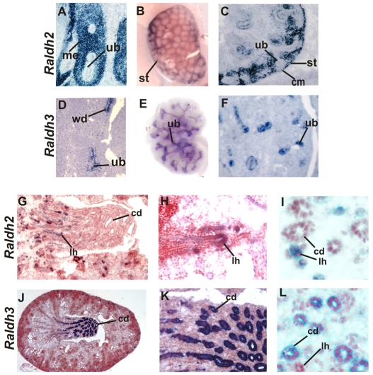
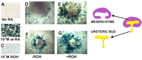
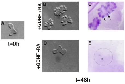
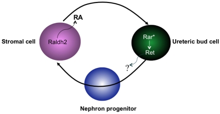
In humans and mice, mutations in the Ret gene result in Hirschsprung's disease and renal defects. In the embryonic kidney, binding of Ret to its ligand, Gdnf, induces a program of epithelial cell remodeling that controls primary branch formation and branching morphogenesis within the kidney. Our previous studies showed that transcription factors belonging to the retinoic acid (RA) receptor family are crucial for controlling Ret expression in the ureteric bud; however, the mechanism by which retinoid-signaling acts has remained unclear. In the current study, we show that expression of a dominant-negative RA receptor in mouse ureteric bud cells abolishes Ret expression and Ret-dependent functions including ureteric bud formation and branching morphogenesis, indicating that RA-receptor signaling in ureteric bud cells is crucial for renal development. Conversely, we find that RA-receptor signaling in ureteric bud cells depends mainly on RA generated in nearby stromal cells by retinaldehyde dehydrogenase 2, an enzyme required for most fetal RA synthesis. Together, these studies suggest that renal development depends on paracrine RA signaling between stromal mesenchyme and ureteric bud cells that regulates Ret expression both during ureteric bud formation and within the developing collecting duct system.


Figures

Fig. 1.
Fig. 1.
Raldh2 and Raldh3 are selectively expressed in cortical stroma and in the ureteric bud during kidney development. (A-C,G-I) Raldh2 expression in a section from an E11 wild-type embryo (A), a wholemount E14 wild-type kidney (B), a section of an E14 wild-type embryonic kidney (C), a section of an E18 wild-type kidney (G), a higher magnification of G (H), and a section through an E18 wild-type embryonic kidney (I). (D-F,J-L) Raldh3 expression in a section of an E11 wild-type embryonic kidney (D), a wholemount E14 wild-type embryonic kidney (E), a section of an E14 wild-type embryonic kidney (F), a section of an E18 wild-type embryonic kidney (J), a higher magnification J (K), and a section of an E18 wild-type embryo (L). cd, collecting duct; cm, cap mesenchyme; lh, Loop of Henle; me, mesenchyme; st, stroma; ub, ureteric bud; wd, Wolffian duct. Magnifications: 40× in I,L; 20× in A,C,D,F,H,K; 10× in G; 5× in B,E,J.
Fig. 2.
Fig. 2.
Renal development depends mainly on Raldh2. (A-F) A Hematoxylin and Eosin (H/E)-stained section through an E12 kidney from a wild-type embryo (A), an E12 Raldh2 mutant embryonic kidney (B), an E14 wild-type kidney (C), an E14 Raldh2 mutant kidney (D), an E14 Raldh3 mutant kidney (E), and an E14 kidney from a Raldh2–/–;Raldh3–/– mutant (F). (G-L) In situ hybridization showing Ret expression in an E12 wild-type kidney (G), an E12 Raldh2 mutant kidney (H), an E14 wild-type kidney (I), an E14 Raldh2 mutant kidney (J), an E14 Raldh3 mutant kidney (K), and an E14 Raldh2–/–;Raldh3–/– compound mutant kidney (L). (M) A wholemount E18 kidney from a wild-type Hoxb7-Gfp transgenic embryo. (N) A vibratome section through an E18 Hoxb7-Gfp wild-type embryo. (O) A wholemount E14 control Hoxb7-Gfp embryonic kidney. (P) A wholemount E18 Hoxb7-Gfp;Raldh2–/– kidney. (Q) A vibratome section through an E18 Hoxb7-Gfp;Raldh3–/– embryonic kidney. (R) A wholemount Hoxb7-Gfp;Raldh3–/– E14 kidney. ki, kidney; ub, ureteric bud. Magnifications: 10× in A-L,O,R; 5× in M,N,P,Q.
Fig. 3.
Fig. 3.
RA is synthesized by both the ureteric bud and mesenchyme in the embryonic kidney. (A-C) LacZ expression in F9-RARE;lacZ reporter cells cultured for 16 hours in medium without added retinoic acid (RA) (A), with 10–8 M at 9-cis RA (B) and with 10–7 M retinol (C), an inactive RA precursor. (D-G) LacZ expression in explants cultured for 16 hours on lawns of F9-RARE;lacZ reporter cells: E11 mesenchyme without a source of vitamin A (retinol) (D), E11 mesenchyme with 10–7 M retinol (E), E11 ureteric buds plated without retinol (F) and E11 ureteric buds with 10–7 M retinol (G). The schematic on the right shows separated metanephric mesenchyme (purple, assayed for lacZ activity in 3D,E) and ureteric bud explants (yellow, assayed for lacZ activity in 3F,G). Magnification: 20× in A-G.
Fig. 4.
Fig. 4.
RA is sufficient for maintaining Ret expression and branching of isolated ureteric buds in the absence of mesenchyme. (A) A wholemount E11 ureteric bud prior to culture. (B) E11 ureteric buds cultured in serum-free medium including Gdnf, with added retinoic acid (RA). (C) In situ hybridization showing Ret expression in ureteric buds cultured for 48 hours in medium containing Gdnf and RA. (D) A wholemount ureteric bud cultured for 48 hours in medium with Gdnf and without added RA. (E) Undetectable Ret expression in an E11 ureteric bud cultured for 48 hours in medium with Gdnf and without RA. Magnification: 20× in A-E.
Fig. 5.
Fig. 5.
Expression of RaraDN in ureteric bud cells inhibits Ret expression and branching in a dose-dependent manner. (A) A lacZ-stained section from a control (Hoxb7-Cre–/–;RaraDNflox/flox;RARE-lacZ) embryo. (B) A lacZ-stained section of an embryo expressing one allele (Hoxb7-Cre–/+;RaraDNflox/+;RARE-lacZ) of the RaraDN transgene. (C) Undetectable lacZ expression in kidneys in an embryo expressing two alleles (Hoxb7-Cre–/+;RaraDNflox/flox;RARE-lacZ) of the RaraDN transgene. (D) A Hematoxylin and Eosin (H/E)-stained section from an E14 Hoxb7-Cre–/–;RaraDNflox/flox control embryonic kidney. (E) An H/E-stained section through a kidney from an Hoxb7-Cre–/+;RaraDNflox/flox embryo. (F) In situ hybridization analysis of a control (Hoxb7-Cre–/–;RaraDNflox/+) embryonic kidney. (G) In situ hybridization analysis of an embryo expressing one allele (Hoxb7-Cre–/+;RaraDNflox/+) of the RaraDN transgene. (H) Undetectable Ret expression in a sectioned kidney from an E14 (Hoxb7-Cre–/+;RaraDNflox/flox embryo) expressing two alleles of the RaraDN transgene. (I) Hoxb7-Gfp expression in a control (Hoxb7-Cre–/–;RaraDNflox/flox) E14 embryonic kidney (J) Reduced branching morphogenesis in a Hoxb7-Cre–/+;RaraDNflox/+;Hoxb7-Gfp E14 kidney from an embryo expressing one allele of the dominant-negative transgene. (K,L) Renal hypoplasia and renal agenesis, respectively, in Hoxb7-Cre–/+;RaraDNflox/flox;Hoxb7-Gfp E14 embryonic kidneys from animals expressing two alleles of dominant-negative RaraDN transgene. The asterisk in (L) denotes renal agenesis. gu, gut; ki, kidney; te, testes; ub, ureteric bud. Magnification: 10× in A-L.
Fig. 6.
Fig. 6.
RA signaling is important for primary ureteric bud formation as well as for branching morphogenesis within the kidney. (A,B) Ureteric bud formation in a wholemount E10.5 control (Hoxb7-Cre–/–;RaraDNflox/flox;Hoxb7-Gfp) embryo (A) and an E10.5 Hoxb7-Cre–/+;RaraDNflox/flox;Hoxb7-Gfp embryo expressing two alleles of the RaraDN transgene (B). (C,D) Gfp expression in an E11.5 control embryo (C) and an E11.5 Hoxb7-Cre–/–;RaraDNflox/flox;Hoxb7-Gfp embryo (D). (E,F) Wholemount in situ hybridization showing Ret expression in the emerging ureteric bud of a control (Hoxb7-Cre–/–;RaraDNflox/flox) E10.5 embryo (E) and an E10.5 Hoxb7-Cre–/+;RaraDNflox/flox;Hoxb7-Gfp embryo (F). The black arrows point to the caudal Wolffian ducts. Magnification: 20× in A-F.
Fig. 7.
Fig. 7.
Model showing Ret regulation in ureteric bud cells by paracrine RA signaling. Secreted retinoic acid (RA) synthesized by Raldh2 in cortical stroma binds to RA receptors in ureteric bud cells and activates Rar-dependent signaling (Rar*). RA-dependent transcription induces Ret expression in ureteric bud cells, either directly or indirectly.

Similar articles

Cited by

References

    1. Batourina E., Gim S., Bello N., Shy M., Clagett-Dame M., Srinivas S., Costantini F., Mendelsohn C. (2001). Vitamin A controls epithelial/mesenchymal interactions through Ret expression. Nat. Genet. 27, 74-78 - PubMed
    1. Batourina E., Tsai S., Lambert S., Sprenkle P., Viana R., Dutta S., Hensle T., Wang F., Niederreither K., McMahon A. P., et al. (2005). Apoptosis induced by vitamin A signaling is crucial for connecting the ureters to the bladder. Nat. Genet. 37, 1082-1089 - PubMed
    1. Bhat P. V., Manolescu D. C. (2008). Role of vitamin A in determining nephron mass and possible relationship to hypertension. J. Nutr. 138, 1407-1410 - PubMed
    1. Blumberg B., Bolado J., Jr, Moreno T. A., Kintner C., Evans R. M., Papalopulu N. (1997). An essential role for retinoid signaling in anteroposterior neural patterning. Development 124, 373-379 - PubMed
    1. Brenner B. M., Mackenzie H. S. (1997). Nephron mass as a risk factor for progression of renal disease. Kidney Int. Suppl. 63, S124-S127 - PubMed

Publication types

MeSH terms