. 2009 Dec;21(12):3839-49.
doi: 10.1105/tpc.109.071639. Epub 2009 Dec 29.

PIN auxin efflux carrier polarity is regulated by PINOID kinase-mediated recruitment into GNOM-independent trafficking in Arabidopsis

Affiliations

PIN auxin efflux carrier polarity is regulated by PINOID kinase-mediated recruitment into GNOM-independent trafficking in Arabidopsis

Jürgen Kleine-Vehn et al. Plant Cell. 2009 Dec.

Abstract

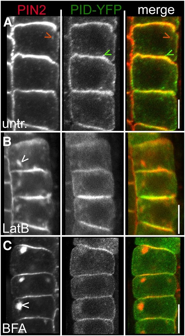
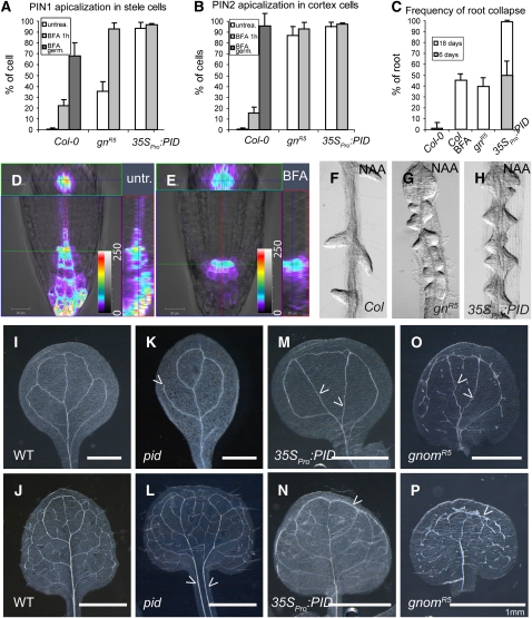
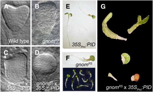
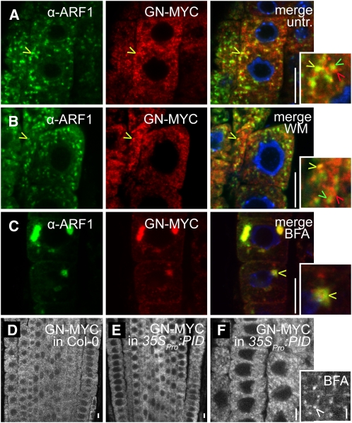
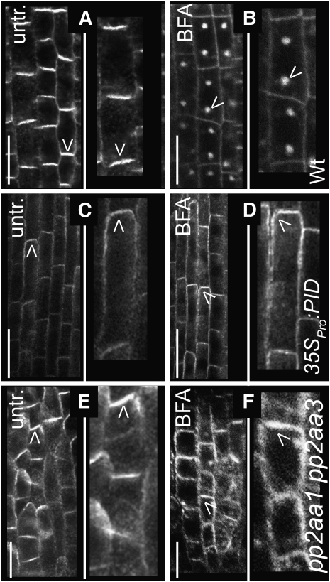
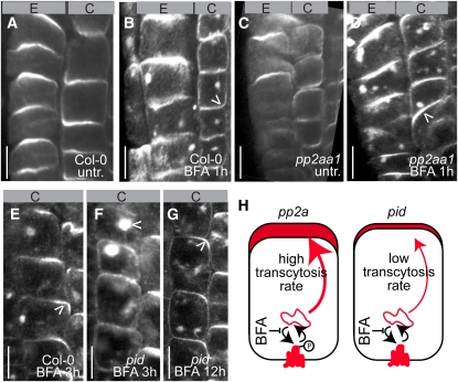
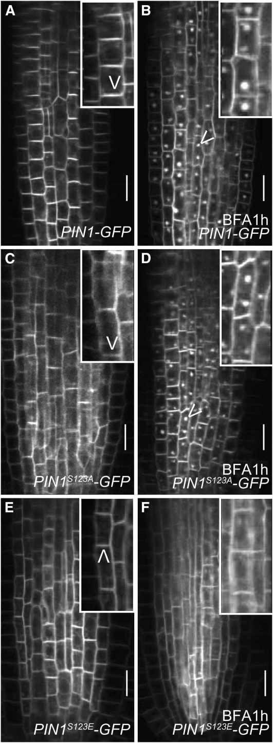
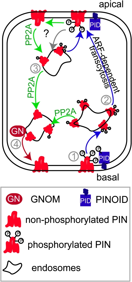
The phytohormone auxin plays a major role in embryonic and postembryonic plant development. The temporal and spatial distribution of auxin largely depends on the subcellular polar localization of members of the PIN-FORMED (PIN) auxin efflux carrier family. The Ser/Thr protein kinase PINOID (PID) catalyzes PIN phosphorylation and crucially contributes to the regulation of apical-basal PIN polarity. The GTP exchange factor on ADP-ribosylation factors (ARF-GEF), GNOM preferentially mediates PIN recycling at the basal side of the cell. Interference with GNOM activity leads to dynamic PIN transcytosis between different sides of the cell. Our genetic, pharmacological, and cell biological approaches illustrate that PID and GNOM influence PIN polarity and plant development in an antagonistic manner and that the PID-dependent PIN phosphorylation results in GNOM-independent polar PIN targeting. The data suggest that PID and the protein phosphatase 2A not only regulate the static PIN polarity, but also act antagonistically on the rate of GNOM-dependent polar PIN transcytosis. We propose a model that includes PID-dependent PIN phosphorylation at the plasma membrane and the subsequent sorting of PIN proteins to a GNOM-independent pathway for polarity alterations during developmental processes, such as lateral root formation and leaf vasculature development.


Figures

Figure 1.
Figure 1.
Subcellular Localization of PIN2 and PID in Root Epidermal Cells. (A) to (C) Confocal images of anti-PIN2 and anti-GFP immunolocalizations. Bars = 10 μm. (A) Pronounced colocalization of PIN2 (left panel/red in the merged image) and PID-YFP (middle panel/green in the merged image) at the plasma membrane, whereas no pronounced colocalization in the endosomes could be detected (indicated by red and green arrowheads for endosomal PIN2 and PID signal, respectively). (B) and (C) Strong intracellular PIN2 localization (depicted by white arrowheads) induced by latrunculin B (B) and BFA (C) treatments but weaker and more dispersed intracellular PID-YFP accumulations.
Figure 2.
Figure 2.
Opposite Actions of GNOM and PID on PIN Polarity and Plant Development. (A) and (B) Apicalization of PIN1 (A) in stele and PIN2 (B) in cortex cells of wild-type, partial loss of GNOM function [gnomR5, labeled gn(R5)], and PID gain-of-function (35SPro:PID) lines. White bars represent the untreated condition, and gray bars illustrate BFA treatment for 1 h (light gray) or germination on medium containing BFA (dark gray). At least 1000 stele and 400 cortex cells for each treatment or genotype (roots, n > 12) were counted. Error bars indicate sd. (C) Frequency of primary root collapse in 35SPro:PID, BFA-treated wild-type plants and the weak gnomR5 allele after 18 d (and after 6 d for 35SPro:PID). Error bars indicate sd (n = 30 seedlings). (D) and (E) Confocal z-stack analysis and subsequent fluorescence intensity profiling (red/yellow denotes high fluorescence intensities and blue/purple denotes low) of the auxin-responsive promoter element DR5revPro:GFP in untreated (D) and BFA-treated (E) seedlings. The central image shows a single medium confocal section, while the top (green) and right (red) insets represent the radial (green line) and longitudinal (red line) distributions of the signal, respectively, giving three-dimensional information of the signal intensity. Under untreated conditions, DR5 signal is the highest in the quiescent center and outermost tier of columella cells (D). By contrast, BFA-dependent inhibition of GNOM function leads to depletion of the response maximum in the root tip and radial expansion of the signal (E). (F) to (H) Synthetic auxin 1-naphthyl acetic acid (NAA) treatment induces enhanced, but spaced, lateral root development in wild-type seedlings (F). By contrast, both gnomR5 (G) and 35SPro:PID (H) mutants display defective primordium spacing and development after NAA treatment. (I) to (P) Vascular development of cotyledons ([I], [K], [M], and [O]) and leaves ([J], [L], [N], and [P]) in wild-type ([I] and [J]), pid ([K] and [L]), 35SPro:PID ([M] and [N]), and gnomR5 ([O] and [P]) backgrounds. Arrows point out vein discontinuity (K), vein polarity defects ([L], [M], and [O]), and enhanced cortical vascularization ([N] and [P]).
Figure 3.
Figure 3.
Genetic Interaction of PID and GNOM. (A) to (D) Patterning defects in mutant embryos compared with wild-type embryos (A). Apical and basal patterning defects in gnomR5 mutant embryos (B). More severe apical-to-basal embryonic patterning defects in 35SPro:PID gnomR5 double mutants (C), sometimes leading to the complete loss of apical basal patterning (D). (E) and (F) Viable 35SPro:PID (E) and gnomR5 (F) seedlings. (G) Strongly affected seedling morphology and growth arrest in 35SPro:PID gnomR5 double mutants, resembling full gnom knockout mutants.
Figure 4.
Figure 4.
PID-Independent GNOM Localization. (A) to (F) Confocal images of anti-ARF1 and anti-MYC immunolocalizations. Bars = 10 μm. (A) to (C) Partial ARF1 (green) and GNOM (GN-MYC) (red) colocalization in untreated seedlings (A) and seedlings treated with wortmannin (B) or BFA (C). Insets display enlarged regions highlighting colocalizing (yellow arrowheads) and noncolocalizing (red and green arrowheads) endosomes. (D) and (E) Similar expression pattern of GNOM (GN-MYC under endogenous promoter) in the wild type (D) and 35SPro:PID (E). (F) Normal subcellular distribution and response to BFA (BFA treatment is shown in the inset) of GNOM-MYC in 35SPro:PID-expressing seedlings. White arrowhead indicates GNOM accumulation in the BFA compartment.
Figure 5.
Figure 5.
BFA-Insensitive PIN Trafficking by Antagonistic PID and PP2A Action. (A) to (F) Confocal images of anti-PIN1 immunolocalizations. Arrowheads indicate the most pronounced localization of endogenous PIN1 at the apical/basal cell side or in BFA compartments. Bars = 10 μm. (A) Basal PIN1 localization in stele cells of untreated wild-type seedlings. (B) Rapid PIN internalization after BFA (50 μM, 1 h) treatment in wild-type seedlings. (C) Apical PIN1 localization in untreated stele cells as a consequence of PID gain of function. (D) Largely BFA-insensitive PIN1 localization in 35SPro:PID, displaying a reduced accumulation in BFA compartments and persistent labeling of the plasma membrane. (E) Preferentially apical PIN1 localization due to partial loss of PP2A function in untreated pp2aa1 pp2aa3 double mutants. (F) Reduced sensitivity to BFA of PIN1 trafficking in pp2aa1 pp2aa3 double mutants.
Figure 6.
Figure 6.
PP2A and PID Modulate the BFA-Induced Transcytosis of PIN Proteins. (A) to (G) Confocal images of anti-PIN2 immunolocalizations. Arrowheads indicate the most pronounced localization of endogenous PIN2 at the apical/basal side of the cell or in BFA compartments. E, epidermal cell files; C, cortical cell files. Bars = 10 μm. (A) to (D) Wild-type seedlings (untreated in [A]) display only weak basal-to-apical transcytosis of PIN2 in cortex cells after 1 h of BFA (50 μM) treatment (B). Enhanced basal-to-apical transcytosis of PIN2 in cortex cells of pp2a mutants (untreated in [C]) after 1 h of BFA (50 μM) treatment (D). (E) to (G) Preferential apical PIN2 localization in lower cortex cells of wild-type seedlings after 3 h of 50 μM BFA incubation (E). Strong PIN2 accumulation in BFA compartments and reduced basal-to-apical transcytosis of PIN2 in cortex cells in pid mutants (F). PIN2 recruitment to the apical side of the cell in lower cortex cells after prolonged BFA treatments (12 h) in the pid mutant background (G). (H) Scheme depicting altered affinity (depicted by thickness of the arrow) of PIN proteins for the apical targeting machinery and subsequent PIN transcytosis rate (depicted at the upper side of the cell) in pp2aa1 and pid mutants. [See online article for color version of this figure.]
Figure 7.
Figure 7.
BFA-Independent PIN Targeting by Phosphorylation-Based Sequence Modification. (A) to (F) Confocal live cell imaging of PIN1-GFP variants. Bars = 10 μm. (A) and (B) PIN1-GFP localization in the root (untreated in [A]) is sensitive to BFA treatment, leading to PIN1-GFP accumulation in BFA compartments (B). (C) and (D) Nonphosphorylated PIN (PIN1S123A-GFP) targeting in untreated (C) and BFA-treated seedlings (D). (E) and (F) Phosphorylation-mimicking PIN1S123E-GFP localization in untreated (E) and BFA-treated seedlings (F), indicating that the phosphorylated PIN proteins preferentially traffic in a BFA-resistant manner.
Figure 8.
Figure 8.
Model of PID and GNOM-Dependent Intracellular PIN Sorting. PID might phosporylate PIN proteins preferentially at the plasma membrane (1). Phosporylated PIN proteins get internalized into endosomes but fail to get sorted to the ARF-GEF GNOM-dependent basal recycling pathway (4). Phosphorylated PIN proteins display an enhanced affinity for a GNOM-independent, but ARF-dependent, apical targeting pathway, eventually leading to basal-to-apical PIN transcytosis (2). PP2AA function can counteract (3) the PID-dependent PIN phosphorylation, leading to GNOM-dependent (GN) basal recycling of PIN proteins (4). [See online article for color version of this figure.]

Similar articles

Cited by

References

    1. Abas, L., Benjamins, R., Malenica, N., Paciorek, T., Wiśniewska, J., Moulinier-Anzola, J.C., Sieberer, T., Friml, J., and Luschnig, C. (2006). Intracellular trafficking and proteolysis of the Arabidopsis auxin-efflux facilitator PIN2 are involved in root gravitropism. Nat. Cell Biol. 8 249–256. - PubMed
    1. Benjamins, R., Quint, A., Weijers, D., Hooykaas, P., and Offringa, R. (2001). The PINOID protein kinase regulates organ development in Arabidopsis by enhancing polar auxin transport. Development 128 4057–4067. - PubMed
    1. Benková, E., Michniewicz, M., Sauer, M., Teichmann, T., Seifertová, D., Jürgens, G., and Friml, J. (2003). Local, efflux-dependent auxin gradients as a common module for plant organ formation. Cell 115 591–602. - PubMed
    1. Chow, C.-M., Neto, H., Foucart, C., and Moore, I. (2008). Rab-A2 and Rab-A3 GTPases define a trans-Golgi endosomal membrane domain in Arabidopsis that contributes substantially to the cell plate. Plant Cell 20 101–123. - PMC - PubMed
    1. Christensen, S.K., Dagenais, N., Chory, J., and Weigel, D. (2000). Regulation of auxin response by the protein kinase PINOID. Cell 100 469–478. - PubMed

Publication types

MeSH terms

Substances