. 2010 Apr;38(6):1950-63.
doi: 10.1093/nar/gkp1190. Epub 2009 Dec 29.

Rearrangement of the RNA polymerase subunit H and the lower jaw in archaeal elongation complexes

Affiliations

Rearrangement of the RNA polymerase subunit H and the lower jaw in archaeal elongation complexes

Sebastian Grünberg et al. Nucleic Acids Res. 2010 Apr.

Abstract

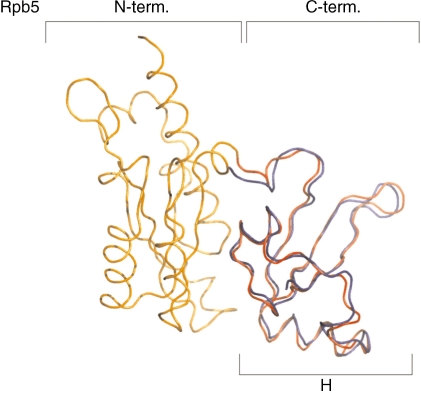
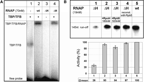
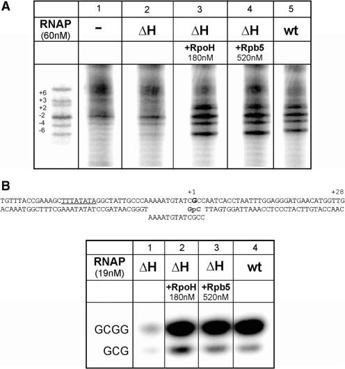
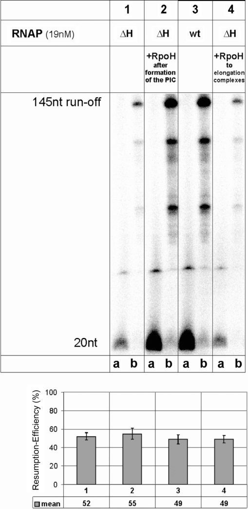
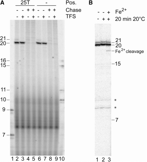
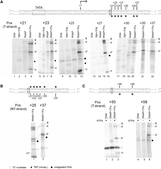
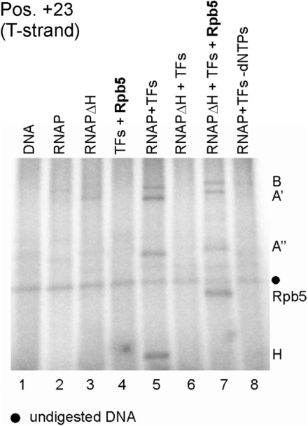
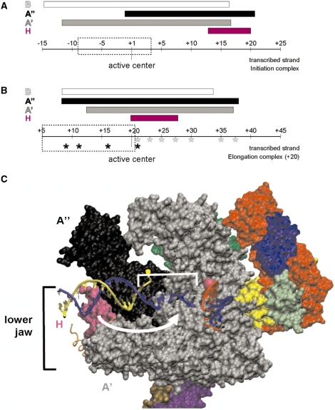
The lower jaws of archaeal RNA polymerase and eukaryotic RNA polymerase II include orthologous subunits H and Rpb5, respectively. The tertiary structure of H is very similar to the structure of the C-terminal domain of Rpb5, and both subunits are proximal to downstream DNA in pre-initiation complexes. Analyses of reconstituted euryarchaeal polymerase lacking subunit H revealed that H is important for open complex formation and initial transcription. Eukaryotic Rpb5 rescues activity of the DeltaH enzyme indicating a strong conservation of function for this subunit from archaea to eukaryotes. Photochemical cross-linking in elongation complexes revealed a striking structural rearrangement of RNA polymerase, bringing subunit H near the transcribed DNA strand one helical turn downstream of the active center, in contrast to the positioning observed in preinitiation complexes. The rearrangement of subunits H and A'' suggest a major conformational change in the archaeal RNAP lower jaw upon formation of the elongation complex.


Figures

Figure 1.
Figure 1.
The C-terminal domains of Rpb5 and RpoH are very similar. Superposition of the tertiary structures of subunits Rpb5 from yeast based on the RNAP II crystal structure (N-terminal domain coloured in orange and the C-terminal domain in red; PDB ID: 1I3Q; 8) and of the archaeal subunit H from S. solfataricus (coloured in blue; PDB ID: 2PMZ; 9) generated using DaliLite (45). The R.M.S. value of this alignment is 1.1 Å.
Figure 2.
Figure 2.
Characterization of promoter recruitment and of run-off transcripts of ΔH RNAP. (A) ΔH RNAP forms stable preinitiation complexes. EMSAs with a probe containing the Pyrococcus gdh promoter (10) were conducted in the presence and absence of TBP, TFB and reconstituted wt or ΔH RNAP as indicated on top of the lanes. The position of the TBP–TFB and TBP–TFB–RNAP complexes are indicated. (B) Eucaryotic Rpb5 can functionally replace RpoH in an archaeal RNAP. The synthesis of a 145 nt run-off transcript from the Pyrococcus gdh promoter was analyzed in standard multiple round transcription assays (‘Materials and Methods’ section) and RNA products were analyzed on 6% denaturing PA gels. Lane 4 shows transcription products synthesized by ΔH RNAP reconstituted with Rpb5. In lanes 2 and 3 subunit H or Rpb 5 were added to transcription reactions. The diagram below the gel shows the mean value of the transcriptional activity for each lane. The quantification was done with the Aida image analyzer software version 3.28.
Figure 3.
Figure 3.
Subunit H is required for open complex formation and initial transcription. (A) RpoH and Rpb5 stimulate open complex formation. Promoter opening at 70°C was analyzed by KMnO4 footprinting as described (16,23). TBP, TFB, TFE and the RpoE′-F complex were present in all reactions. Additional components added to the reactions are indicated on top of the gel. (B) Abortive transcription from a preopened heteroduplex. The ability of ΔH and of wt RNAP to synthesize a 3 or 4 nt abortive transcript in the presence of TBP and TFB was analyzed on a preopened bubble mimicking an open complex (see figure on top of the gel). The reaction was dinucleotide (GpC) primed and was analyzed on a 24% PA gel. Subunits H and Rpb5 were added to transcription reactions as indicated.
Figure 4.
Figure 4.
Stalled ΔH RNAPs can efficiently elongate to the run-off. Ternary complexes were stalled at +20, washed and the reactions divided into two aliquots. One aliquot was untreated (lane a of each panel), the other was chased by the addition of a complete set of non-labeled NTPs for 3 min at 70°C (lane b in each panel), and both were then analyzed on a 20% polyacrylamide gel. In lanes 1, 2 and 4, the ability of the ΔH and wt enzyme to synthesize a 20-nt stalled transcript in the presence and absence of subunit H was analyzed. Reactions were chased by the addition of a complete set of NTPs not containing radioactivity and the synthesis of the 145-nt run-off product was analyzed. The RNA products of intermediate size are caused by specific pausing sites for RNAP. The quantification below shows the mean value of the transcriptional activity of the transcript in ternary complexes stalled at position +20 relative to the 145-nt run-off product.
Figure 5.
Figure 5.
Resumption of stalled complexes and Fe2+ cleavage indicate that EC20 complexes are not backtracked. (A) Complexes stalled at the gdh promoter at position +20 on a template with and without aryl azide derivatization at position +25 of the template strand (25T; lanes 2, 3 and 6, 7) were challenged after incubation for 5 min at 70°C with a complete set of NTPs (chase) for 2 min in the presence and absence of TFS as indicated on top of the figure. Lanes 1 and 10 contained RNA size markers. (B) Stalled immobilized EC20 were extensively washed in the absence of Mg2+, as indicated in ‘Materials and Methods’ section, and incubated for 20 min at 70°C. Reactions were then cooled to 20°C, and Fe2+ and DTT were added to initiate the cleavage reaction. The ∼18-nt Fe2+ cleavage product contains a 3′ phosphate causing as slightly higher electrophoretic mobility (39). The weak 21-nt RNA band is due to misincorporation at the G-residue of template DNA.The asteriks indicate non-specific bands.
Figure 6.
Figure 6.
Mapping of RNAP subunits cross-linked to the gdh promoter DNA in stalled elongation complexes. (A) Processivity of ECs stalled at +20 is not affected by TFS. Increasing amounts of purified TFS were added to stalled ECs (lanes 4–9) and complexes were chased with a complete set of NTP after incubation in the absence (lane 3) and presence of TFS (lane 8). The amount of non-chaseable RNA in complexes is insignificant in both reactions and the amount of synthesized run-off transcript ∼ the same indicating that the stalled complexes were not backtracked in the absence of TFS. As expected, the 21-nt RNA most likely generated by misincorporation of an unpaired nt was removed upon TFS treatment. (B) Photocross-linking of proteins in a ternary complex stalled at position +20. The gdh promoter template spans bp −39 to +66 relative to the transcription start site (bent arrow). TATA box and the stalling site +20 are boxed. Locations of photoactivatable labels (4-azidophenacyl bromide coupled to a phosphorothioate modification in the DNA backbone) are indicated by asterisks. RNAP purified from Pyrococcus cells was used to stall elongation complexes at +20 as described earlier (6,27). Cross-linked subunits of the stalled complexes were analyzed via 4–20% SDS-PAGE. The position of the photoactive cross-linking site in the transcribed strand is indicated above the gels. Proteins present in the individual reactions are specified on top of the gels. Radiolabeled RNAP subunits were identified by their relative electrophoretic mobility and are indicated at the right-hand side of the gels. Dots indicate non-specific signals derived from undigested DNA, whereas triangles indicate non-specific TBP cross-links. (C) Cross-linking of RNAP subunits in stalled elongation complexes to the non-transcribed strand. Positions analyzed are indicated by asterisks above the sequence. No cross-linking of H was detectable at any of the tested positions except of position +25, where a very faint band suggests weak cross-linking of H. To exemplify this, positions +25 and +37 are shown. (D) H interacts with DNA close to the active center also in late elongation complexes. Mature ECs, stalled at position +45 were cross-linked to position +50 and +58 on the T-strand as described in (B). The diamond marks auto-radiolabeled S7 nuclease. Part of the DNA sequence is shown at the top of the panel.
Figure 7.
Figure 7.
Rpb5 incorporated into the archaeal RNAP cross-links at the same position in elongation complexes as H. Archaeal ΔH RNAP complemented in transcription reactions with Rpb5 was cross-linked in ECs stalled at position +20 (lane 7). Reactions contained transcription factors TFB and TBP (TFs), RNAP purified from Pyrococcus cells (lanes 2, 5 and 8), reconstituted ΔH RNAP and subunit Rpb5 as indicated on top of the lanes. The template DNA was derivatized with APB at position +23. Note that the presence of Rpb5 is essentially required for the formation of cross-linkable stalled ECs by the ΔH RNAP (compare lanes 6 and 7). Subunits of reconstituted ΔH RNAP run at higher molecular weight because they are His-tagged.
Figure 8.
Figure 8.
Mapping of archaeal RNAP and eukaryotic polII subunits cross-linked to an elongation scaffold. The templates for this assay were assembled essentially as described (29) but contained an azidophenacylated phosphorothioate substitution at positions +6 or +15, respectively, with adjacent internal radiolabel. (A) Sequence of the template in the final assembly. RNA is marked in bold gray letters, position of the cross-linker is indicated with asterisks, and internal radiolabels are highlighted in bold letters. (B) Cross-linking of archaeal RNAP subunits to positions +6 (left) and +15 (right). ECs were formed as described in ‘Materials and Methods’ section in the presence of heparin as non-specific competitor. Lanes 1, 3 and 4 contained TFE, which cross-linked non-specifically to both templates as described (6). Radiolabeled RNAP subunits and TFE were identified by their relative electrophoretic mobility and are indicated at the right-hand side of the gels. Note that subunit H cross-links only to position +6 and not to position +15. (C) Cross-linking of polII subunits to positions +6 (left) and +15 (right) on an elongation scaffold. Control lanes 1 and 5 contained the template without polII. Triangles above lanes 2–4 and 6–8 indicate an increase of polII from 36 to 110 nM polII in the reaction. As described in (B), po lII subunits were identified by their mobility by 4–19 % SDS-PAGE. As in (B), diamonds mark auto-radiolabeled S7 nuclease, while dots specify undigested DNA. (D) Eukaryotic polII is active on elongation scaffolds. Lane 1 shows RNA products synthesized in a standard in vitro transcription assay containing 46 nM archaeal nat RNAP and Pyrococcus gdh promoter DNA. Reactions analyzed in lanes 2–4 contained increasing amounts (36–110 nM) of polII and the elongation scaffold shown in (A) as template (for details see ‘Materials and Methods’ section).
Figure 9.
Figure 9.
Schematic summary of the repositioning of H and A′′ in elongation complexes. The range of protein/DNA interaction of RNAP subunits H (magenta), A′ (black), A′ (gray) and B (white) is symbolized by bars. The position of the RNAP’s active center is highlighted and the extension of the transcription bubble (27) is marked by dashed boxes. (A) Interactions of archaeal RNAP subunits with the transcribed DNA-strand in the PIC. H marks the far downstream end of the complex (based on the results published by ref. 17). (B) Schematic representation of cross-links of archaeal RNAP in a ternary complex stalled at +20. Cross-linking from position +9 to +21 was analyzed previously (6), and those derivatized sites are labeled by black asterisks. Cross-linkers from position +21 to +37 from this study are indicated with gray asterisks. Note that H is localized in close proximity to the active center and that the upstream boundary of A′′ is extended by ∼10 nt in stalled ECs. (C) Model for the path of DNA relative to RNAP in the archaeal elongation complex. The S. shibatae RNAP structure (PDB ID: 2WAQ; 13) was aligned with eukaryotic RNAP II in an EC (PDB ID: 2E2H; 35) using C-alpha coordinate ‘Iterative Magic Fit’ from Swiss PDBViewer 4.01 (54). R.M.S. deviation of the aligned structures was 1.55 Å for 1786 atoms. The RNAP II EC DNA and RNA coordinates were then merged with the archaeal RNAP structure coordinates and rendered concurrently using VMD 1.8.6 (54), revealing a good fit with few clashes. RNAP subunit colors are as used in (9), except for subunits G (darker green, obscured) and Rpo13 (orange trace) that were not part of the structure solved by (9). Subunit B was removed to allow visualization of the nucleic acids. The DNA transcribed and non-transcribed strands are blue and yellow, respectively, and the RNA is red. The active site is indicated by a pink sphere, and represents the +1 position in the EC. +1 to +10 of the T strand is bracketed, and the white arrow suggests the conformational change that subunit H (in magenta) would need to make to be cross-linked by aryl azide derivatizations within this region.

Similar articles

Cited by

References

    1. Hausner W, Wettach J, Hethke C, Thomm M. Two transcription factors related with the eucaryal transcription factors TATA-binding protein and transcription factor IIB direct promoter recognition by an archaeal RNA polymerase. J. Biol. Chem. 1996;271:30144–30148. - PubMed
    1. Bell SD, Jackson P. The role of transcription factor B in transcription initiation and promoter clearance in the Archaeon Sulfolobus acidocaldarius. J. Biol. Chem. 2000;275:12934–12940. - PubMed
    1. Hanzelka BL, Darcy TJ, Reeve JN. TFE, an archaeal transcription factor in Methanobacterium thermoautotrophicum related to eucaryal transcription factor TFIIEalpha. J. Bacteriol. 2001;183:1813–1818. - PMC - PubMed
    1. Bell SD, Brinkman AB, van der Oost J, Jackson SP. The archaeal TFIIEalpha homologue facilitates transcription initiation by enhancing TATA-Box recognition. EMBO Rep. 2001;2:133–138. - PMC - PubMed
    1. Meinhart AJ, Blobel J, Cramer P. An extended winged helix domain in general transcription factor E/IIEalpha. J. Biol. Chem. 2003;278:48267–48274. - PubMed

Publication types

MeSH terms