. 2010 Feb;16(2):338-48.
doi: 10.1261/rna.1779810. Epub 2009 Dec 29.

The nuclear experience of CPEB: implications for RNA processing and translational control

Affiliations

The nuclear experience of CPEB: implications for RNA processing and translational control

Chien-Ling Lin et al. RNA. 2010 Feb.

Abstract

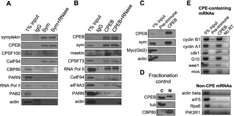
CPEB is a sequence-specific RNA binding protein that promotes polyadenylation-induced translation in early development, during cell cycle progression and cellular senescence, and following neuronal synapse stimulation. It controls polyadenylation and translation through other interacting molecules, most notably the poly(A) polymerase Gld2, the deadenylating enzyme PARN, and the eIF4E-binding protein Maskin. Here, we report that CPEB shuttles between the nucleus and cytoplasm and that its export occurs via the CRM1-dependent pathway. In the nucleus of Xenopus oocytes, CPEB associates with lampbrush chromosomes and several proteins involved in nuclear RNA processing. CPEB also interacts with Maskin in the nucleus as well as with CPE-containing mRNAs. Although the CPE does not regulate mRNA export, it influences the degree to which mRNAs are translationally repressed in the cytoplasm. Moreover, CPEB directly or indirectly mediates the alternative splicing of at least one pre-mRNA in mouse embryo fibroblasts as well as certain mouse tissues. We propose that CPEB, together with Maskin, binds mRNA in the nucleus to ensure tight translational repression upon export to the cytoplasm. In addition, we propose that nuclear CPEB regulates specific pre-mRNA alternative splicing.


Figures

FIGURE 1.
FIGURE 1.
CPEB is a nuclear-cytoplasmic shuttling protein. (A) Left: Western blot of Xenopus oocyte lysate demonstrated the specificity of CPEB antibody used in this study. Right: Nuclei and cytoplasms from oocytes of different stages were manually separated and probed on Western blots for CPEB and tubulin. (B) Stage VI oocytes were treated with 200 nM leptomycin B overnight; nuclei and cytoplasms were then manually separated and probed for CPEB, tubulin as a cytoplasmic marker, and histone H4 as a nuclear marker. (C) MEFs were transfected with CPEB-HA, some of which were then treated with 10 nM LMB for 5 h. The HA epitope was located by indirect immunofluorescence. (D) Lampbrush chromosomes were prepared and immunostained for symplekin and CPEB. Some preparations were treated with RNase before immunolocalization for CPEB. The chromosomes were also stained with DAPI.
FIGURE 2.
FIGURE 2.
CPEB nuclear localization domain. (A) Diagram of deletion mutant constructs of CPEB. PEST refers to a domain rich in proline, glutamic acid, serine, and threonine that is thought to be involved in protein destruction; RRM refers to RNA recognition motif, and ZF refers to zinc finger. (B) Immunocytochemistry of 3T3 cells expressing CPEB-HA full-length or deletions illustrated in panel A. (C) The nucleus-cytoplasm localization was quantified using an arbitrary score; this scoring system was used to analyze the relative localization of the CPEB proteins shown in panel B. Histograms of these data are presented in D; the numbers atop the bars refer to the total number of cells examined.
FIGURE 3.
FIGURE 3.
Requirement for CPEB nuclear localization and RNA binding. (A) CPEB deletion mutants lacking regions between residues 206 and 309 were HA tagged, transfected into NIH 3T3 cells, incubated in the presence of LMB, and analyzed as in Fig. 2. Residues 297–307 were necessary for nuclear localization; each of the 11 residues in this region was changed to alanine and the nuclear localization examined as above. In each case, single alanine substitutions had no effect on nuclear localization. (B) HEK 293T cells were infected with HA-tagged CPEB or CPEB Δ297–307. An extract was then prepared, supplemented with the CPE-containing cyclin B1 3′ UTR, UV irradiated, and subjected to HA immunoprecipitation. The proteins were then analyzed by Western blot for HA (upper panel, two bands are evident; the higher one was likely generated from an upstream cryptic transcription start site of the C-pOZ vector.) and by autoradiography for proteins made radioactive by label transfer (lower panel).
FIGURE 4.
FIGURE 4.
CPEB contains two redundant NESs in the N-terminal half of the protein. (A) When both of the NESs were mutated, CPEB accumulated in the nucleus independently of LMB treatment. (B) An alignment shows these two NESs are conserved among species. The critical leucine/isoleucine residues are also conserved in Drosophila orb1, but not other CPEB family proteins of any species (not shown).
FIGURE 5.
FIGURE 5.
CPEB is a component of the nuclear RNA processing machinery. (A) Symplekin was immunoprecipitated in the absence or presence of RNase A from ∼250 LMB-treated hand-isolated oocyte nuclei. A similar number was mock precipitated with nonspecific IgG. The precipitates were probed on Western blots for the proteins noted in the figure. Actin served as a negative control; 1% of the extract was also applied directly to the gel without immunoprecipitation. (B) Similar to panel A except that CPEB was immunoprecipitated from the nuclear extracts. (C) Oocytes were injected with mRNA encoding myc-tagged Gld2; following overnight culture, the nuclei were isolated and subjected to CPEB immunoprecipitation as in panel B and probed for the proteins noted in the figure. (D) Fractionation control from oocytes used in panels A–C and E; tubulin, a cytoplasmic protein, is entirely cytoplasmic, while CBP80, a nuclear protein, is entirely nuclear. (E) CPEB was immunoprecipitated from oocyte nuclei as before; the RNA was extracted from the precipitates and subjected to RT-PCR for the RNAs noted in the figure.
FIGURE 6.
FIGURE 6.
The nuclear experience of CPE does not mediate mRNA nuclear export or cytoplasmic deadenylation. (A) Diagram of experiment procedure for comparison of RNA export between CPE-containing and CPE-lacking luciferase mRNA. (B) Cytoplasmic luciferase mRNA levels following plasmid injection as determined by radioactive semiquantitative RT-PCR (upper panel). The relative mRNA levels are graphed in the lower panel. (C) Diagram of experiment procedure for comparison of deadenylation between cytoplasm-injected and nucleus-injected cyclin B1 mRNA. (D) Deadenylation assay. A radiolabled and polyadenylated partial cyclin B1 mRNA was injected into the nucleus or cytoplasm of oocytes; after overnight incubation, the cytoplasmic fraction was collected for RNA extraction and analysis on a denaturing polyacrylamide gel. (E) Deadenylation assay of CPE-containing RNA. Oocytes were injected with in vitro transcribed RNA or plasmid DNA; RNA collected over several hours was analyzed by ligation-mediated PAT assay (see Materials and Methods). Lower panels are ethidium bromide stained agarose gels showing RT-PCR products of cyclin B1 RNA; cyclin B1 mRNA started to accumulate ∼3 h after injection in the nucleus and ∼6 h in the cytoplasm. Note that because the RT-PCR does not distinguish endogenous from ectopic cyclin B1 3′ UTR, a band is present in the cytoplasmic fraction of noninjected oocytes.
FIGURE 7.
FIGURE 7.
The nuclear experience of CPE-containing mRNAs mediates tight translational repression. (A) Diagram of experimental procedure for comparing CPE-dependent translational repression with or without the nucleus experience. (B) Time course of translational efficiency of reporters (luciferase activity/RNA) containing or lacking CPEs derived from plasmid DNA-injected oocytes (top). Time course of translational efficiency of the constructs noted above that were synthesized in vitro and then injected into the cytoplasms of oocytes (bottom). (C) Comparison of the translational efficiencies from panel B of plasmid-injected nuclei versus RNA-injected cytoplasm (dark gray bars). Also shown is a comparison of the translational efficiencies between RNA-injected nuclei versus cytoplasm (light gray bars). The RNA was collected 12 to 16 h after injection, and the translation efficiency was determined as in B. The bars represent the fold difference of translational efficiency of RNA lacking the CPE (mt) divided by that of RNA containing the CPE (WT). Statistical analysis was by a one-tailed paired t-test.
FIGURE 8.
FIGURE 8.
CPEB mediates alternative pre-mRNA splicing. (A) RT-PCR (dCTP-[α-P32] incorporation) analysis of exons 33–36 of the collagen 9a1 mRNA from three different WT and CPEB KO MEF lines. (B) RT-PCR analysis of collagen 9a1 mRNA exons 33–36 from different tissues of WT and CPEB KO mice.
FIGURE 9.
FIGURE 9.
Model of CPEB-mediated translational control. A CPE-containing RNA is recognized by a CPEB and Maskin-containing protein complex in the nucleus either co-transcriptionally or soon after transcription is complete. After export from the nucleus, a cytoplasmic RNP complex is assembled that includes the poly(A) polymerase Gld2 and the deadenylating enzyme PARN. PARN is expelled from the complex upon progesterone-induced and aurora A-catalyzed CPEB phosphorylation; Gld2 then elongates the poly(A) tail by default. Maskin is phosphorylated at this time. These events, as well as its association with an embryonic poly(A) binding protein (not shown), lead to the replacement of Maskin for eIF4G on eIF4E. As a consequence, translation is activated. The association of eIF4A3 with the cytoplasmic complex is conjectural.

Similar articles

Cited by

References

    1. Barnard DC, Ryan K, Manley JL, Richter JD. Symplekin and xGLD-2 are required for CPEB-mediated cytoplasmic polyadenylation. Cell. 2004;119:641–651. - PubMed
    1. Bouvet P, Wolffe AP. A role for transcription and FRGY2 in masking maternal mRNA within Xenopus oocytes. Cell. 1994;77:931–941. - PubMed
    1. Braddock M, Thorburn AM, Chambers A, Elliott GD, Anderson GJ, Kingsman AJ, Kingsman SM. A nuclear translational block imposed by the HIV-1 U3 region is relieved by the Tat-TAR interaction. Cell. 1990;62:1123–1133. - PubMed
    1. Brogna S, Wen J. Nonsense-mediated mRNA decay (NMD) mechanisms. Nat Struct Mol Biol. 2009;16:107–113. - PubMed
    1. Cao Q, Richter JD. Dissolution of the maskin-eIF4E complex by cytoplasmic polyadenylation and poly(A)-binding protein controls cyclin B1 mRNA translation and oocyte maturation. EMBO J. 2002;21:3852–3862. - PMC - PubMed

Publication types

MeSH terms