. 2010 Feb 26;285(9):6826-34.
doi: 10.1074/jbc.M109.061846. Epub 2009 Dec 29.

TDP-43 is a developmentally regulated protein essential for early embryonic development

Affiliations

TDP-43 is a developmentally regulated protein essential for early embryonic development

Chantelle F Sephton et al. J Biol Chem. .

Erratum in

  • J Biol Chem. 2010 Dec 3;285(49):38740

Abstract

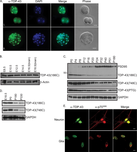
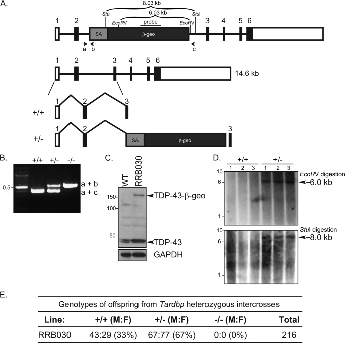
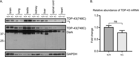
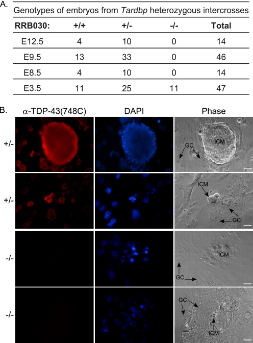
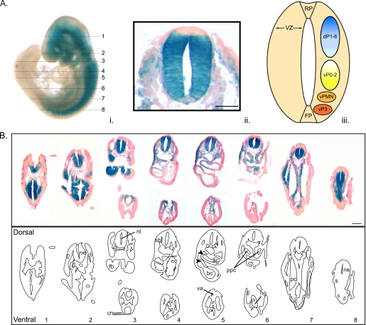
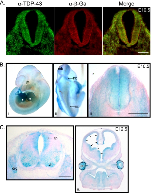
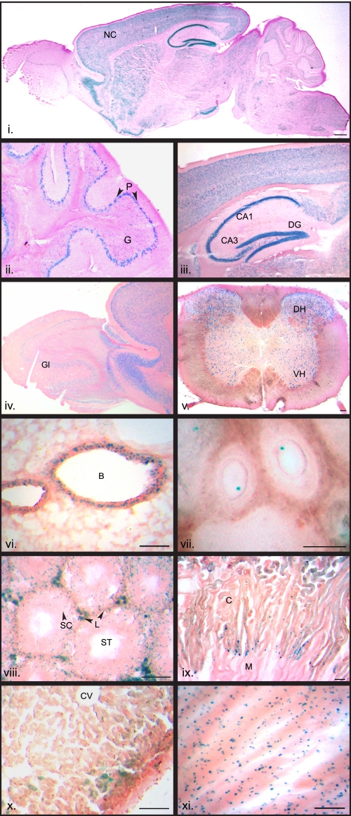
TDP-43 is a DNA/RNA-binding protein implicated in multiple steps of transcriptional and post-transcriptional regulation of gene expression. Alteration of this multifunctional protein is associated with a number of neurodegenerative diseases including amyotrophic lateral sclerosis and frontotemporal lobar degeneration with ubiquitin positive inclusions. Whereas a pathological link to neurodegenerative disorders has been established, the cellular and physiological functions of TDP-43 remain unknown. In this study, we show that TDP-43 is a nuclear protein with persistent high-level expression during embryonic development and with progressively decreased protein levels during postnatal development. In mice where the TDP-43 gene (Tardbp) was disrupted using a gene trap that carries a beta-galactosidase marker gene, heterozygous (Tardbp(+/-)) mice are fertile and healthy, but intercrosses of Tardbp(+/-) mice yielded no viable homozygotic null (Tardbp(-/-)) mice. Indeed, Tardbp(-/-) embryos die between 3.5 and 8.5 days of development. Tardbp(-/-) blastocysts grown in cell culture display abnormal expansion of their inner cell mass. The pattern of beta-galactosidase staining at E9.5 Tardbp(+/-) embryos is predominantly restricted to the neuroepithelium and remains prominent in neural progenitors at E10.5-12.5. TDP-43 is detected in spinal cord progenitors and in differentiated motor neurons as well as in the dorsal root ganglia at E12.5. Beta-galactosidase staining of tissues from adult Tardbp(+/-) mice shows widespread expression of TDP-43, including prominent levels in various regions of the central nervous system afflicted in neurodegenerative disorders. These results indicate that TDP-43 is developmentally regulated and indispensible for early embryonic development.


Figures

FIGURE 1.
FIGURE 1.
Expression of TDP-43 during development. A, embryos (i), two- and six-cell stage, and (ii) blastocyst stage, E3.5, were harvested from superovulated female WT mice from intercrossed mating. Embryos were incubated with anti-TDP-43(PTG), followed by Alexa Fluor 488 (green) and stained with 4′,6-diamidino-2-phenylindole (DAPI) (blue). Scale bars represent 100 μm. B–D, whole embryos (E8.5–14.5) and the brains from embryos (E16.5 and E18.5) and postnatal mice were harvested at the indicated times and proteins were extracted. B, Western blot showing protein expression of TDP-43 in the developing embryo using anti-TDP-43 (186C). Levels of β-Actin were used as a loading control. C, Western blot of brain lysates from postnatal mice probed with anti-TDP-43(186C), anti-TDP-43(748C), commercially available anti-TDP-43(PTG) and anti-PSD95. D, Western blot of brain lysates from E18.5, P0, P180, and P330 using anti-TDP-43(186C) and TDP-43(748C). Levels of glyceraldehyde-3-phosphate dehydrogenase (GAPDH) were used as a loading control. E, mouse primary cortical neurons and glia stained with anti-TDP-43(PTG) and anti-p70S6K, followed by Alexa Fluor 488 (green) and Alexa Fluor 546 (red). Scale bars represent 10 μm.
FIGURE 2.
FIGURE 2.
Generation of TDP-43 knock-out mouse. A, schematic of the gene trap in Tardbp. Numbered boxes represent exons. The mutated allele yields an in-frame fusion transcript with exons 1–2 of Tardbp and β-geo. Oligonucleotide primers a, b, and c are indicated. SA, splice acceptor. B, genotyping of E3.5 embryos by PCR. C, Western blot showing expression of the WT TDP-43 protein in ES cells and expression of the TDP-43-β-geo fusion protein in targeted ES cells using an N-terminal TDP-43 (750C) antibody. Glyceraldehyde-3-phosphate dehydrogenase (GAPDH) was used as a loading control. D, identification of a single gene trap insertion using Southern blot. Genomic DNA was digested with EcoRV or StuI and hybridized with a 32P-labeled neomycin phosphotransferase II probe and the predicted ∼6- or ∼8-kb band, respectively, was detected in Tardbp+/− mice only. E, genotyping results of offspring from intercrossed Tardbp+/− mice.
FIGURE 3.
FIGURE 3.
Expression profile of TDP-43 in Tardbp+/− mice. A, Western blot showing protein expression of TDP-43 in Tardbp+/− and WT (Tardbp+/+) mice using the C-terminal TDP-43(748C) antibody and enhanced chemiluminescence (lower panels) or 125I-labeled anti-rabbit secondary antibody (upper panel). B, quantitative PCR analysis of TDP43 mRNA levels in WT and heterozygous mice using the comparative threshold cycle method relative to U36B as an internal control. Data were analyzed by unpaired Student's t test and are represented as relative to WT mRNA (n = 3; ns, not significant). Error bars represent the mean ± S.E.
FIGURE 4.
FIGURE 4.
TDP-43 is necessary for expansion of the inner cell mass. A, genotyping results of embryos collected at 12.5, 9.5, 8.5, and 3.5 dpc from intercrossed Tardbp+/− mice. B, blastocysts at E3.5 were grown in vitro for 7 days. Outgrowths were incubated with anti-TDP-43(748C), followed by Alexa Fluor 546 (red). Nuclei were stained with 4′,6-diamidino-2-phenylindole (DAPI) (blue). WT and heterozygous embryos displayed expansion of the ICM and giant trophoblasts (GC), whereas homozygous embryos had no ICM expansion. Scale bars represent 100 μm.
FIGURE 5.
FIGURE 5.
TDP-43 expression in the developing mouse embryo. β-Galactosidase staining of a Tardbp+/− embryo at E9.5. A (i), whole mount embryo, numbers indicate corresponding sections shown in 5B. (ii) Transverse section counterstained with nuclear fast red showing X-gal staining in the neuroepithelium of the neural tube. Scale bar represents 100 μm. (iii) Schematic of neuroepithelium indicating the domains for neuronal progenitors. Dorsal progenitors (dP) are indicated as dP1–6. Ventral progenitors (vP) vP0-p3 give rise to interneurons subtypes, vPMN is the source of motor neurons. The floor plate position (FP), an important source of Shh proteins, and the roof plate (RP), a source of bone morphogenetic proteins, are shown (adapted from Refs. and 31). B, transverse sections of whole embryo in series. There is strong staining in the neuroepithelium (ne) of the neural tube and to a lesser extend the first branchial arch (fb), second branchial membrane (sb), atrial heart chamber (ac), vitelline artery (va), bulbus cordis heart region (bc), left and right pericardio-peritoneal canal (ppc), hindgut (hg), and somite (s). Also annotated are the neural folds in caudal neuropore (cn), neural lumen (nl), and peritoneal cavity (pc). The developing heart is indicated by arrows. Scale bars represent 200 μm.
FIGURE 6.
FIGURE 6.
TDP-43 expression in spinal cord and telencephalon progenitor cells. A, cross-section of Tardbp+/− spinal cord showing TDP-43 and β-galactosidase immunostaining at E10.5. B, X-gal staining of a Tardbp+/− embryo at E10.5. (i) Whole mount embryo, asterisks indicate developing limbs. (ii) Dorsal view, showing staining in spinal cord (sc), midbrain (mb), and hindbrain (hb). (iii) Transverse sections of spinal cord showing X-gal staining in the neuroepithelium. C (i), transverse sections of spinal cord of Tardbp+/− at E12.5 showing X-gal staining in spinal cord progenitors (sp), dorsal root ganglia (drg), and in motor neurons of the ventral horn (vh) and (ii) coronal section of the developing brain showing prominent X-gal staining in progenitors (indicated by arrows) and eye (e). X-gal-stained sections were all counterstained with nuclear fast red. Scale bars for A, B, and Ci represent 10 μm and for Cii, 200 μm.
FIGURE 7.
FIGURE 7.
TDP-43 expression in adult tissues. β-Galactosidase expression in adult Tardbp+/− mice was observed in (i) whole brain with prominent staining in the neocortex (NC), (ii) cerebellar cortex staining in the granular cell layer (G) and Purkinje cells (P), and (iii) in the hippocampus (shown are granular dentate gyrus (DG) as well as CA1 and CA3 regions). There is also distinct staining in the (iv) olfactory bulb glomerulus (Gl). Staining is observed throughout the (v) spinal cord mostly in the dorsal horn (DH) and to a lesser extent the ventral horn (VH). Staining is in the bronchiole (B) region of the (vi) lung and throughout the (vii) ovary, particularly in the oocytes, as well as in the (viii) testis, at the periphery of the seminiferous tubules (ST) where Sertioli cells (SC) are found and with some interstitial staining of Leydig (L) cells. Staining in (ix) kidney is concentrated in the (C) cortical region (shown is the medulla (M)). Moderate levels of expression were in the (x) liver (shown is the central vein (CV)) and (xi) heart. Scale bars represent 100 μm.

Similar articles

Cited by

References

    1. Wang H. Y., Wang I. F., Bose J., Shen C. K. (2004) Genomics 83, 130–139 - PubMed
    1. Ayala Y. M., Pantano S., D'Ambrogio A., Buratti E., Brindisi A., Marchetti C., Romano M., Baralle F. E. (2005) J. Mol. Biol. 348, 575–588 - PubMed
    1. Dreyfuss G., Matunis M. J., Piñol-Roma S., Burd C. G. (1993) Annu. Rev. Biochem. 62, 289–321 - PubMed
    1. Buratti E., Baralle F. E. (2008) Front. Biosci. 13, 867–878 - PubMed
    1. Ou S. H., Wu F., Harrich D., García-Martínez L. F., Gaynor R. B. (1995) J. Virol. 69, 3584–3596 - PMC - PubMed

Publication types

Substances

LinkOut - more resources