

Oxidative stress-specific interaction between FANCD2 and FOXO3a
- PMID: 20040763
- PMCID: PMC2830760
- DOI: 10.1182/blood-2009-07-234385
Oxidative stress-specific interaction between FANCD2 and FOXO3a
Abstract
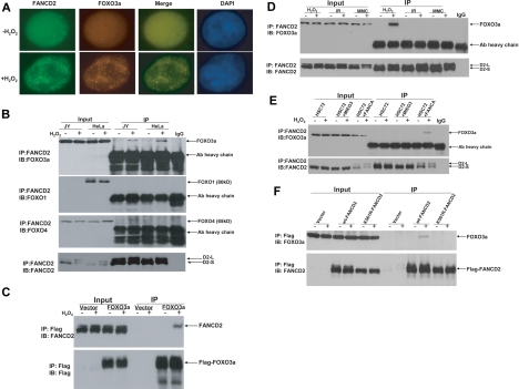
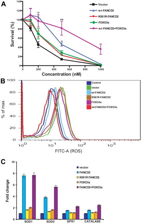
The molecular pathway by which Fanconi anemia (FA) proteins function in oxidative stress response has not been defined. Here we report the functional interaction of the FA protein Fanconi anemia complementation group D2 (FANCD2) and the forkhead transcription factor forkhead box O 3a (FOXO3a). FOXO3a colocalized with FANCD2 foci in response to oxidative stress. The FANCD2-FOXO3a complex was not detected in cells deficient for the FA core complex component FANCA but could be restored in corrected cells. Consistent with this, a nonmonoubiquitinated FANCD2 mutant failed to bind FOXO3a. Although both mitomycin C and ionizing radiation induced FANCD2 monoubiquitination, neither could induce the association of FANCD2 and FOXO3a. Overexpression of FOXO3a reduced abnormal accumulation of reactive oxygen species, enhanced cellular resistance to oxidative stress, and increased antioxidant gene expression in corrected but not mutant FA-D2 cells. The novel oxidative stress response pathway identified in this study, in which FANCD2 and FOXO3a converge, probably contributes to cellular antioxidant defense.
Figures
Similar articles
-
Fancd2 is required for nuclear retention of Foxo3a in hematopoietic stem cell maintenance.J Biol Chem. 2015 Jan 30;290(5):2715-27. doi: 10.1074/jbc.M114.619536. Epub 2014 Dec 12. J Biol Chem. 2015. PMID: 25505262 Free PMC article.
-
Concomitant inactivation of foxo3a and fancc or fancd2 reveals a two-tier protection from oxidative stress-induced hydrocephalus.Antioxid Redox Signal. 2014 Oct 20;21(12):1675-92. doi: 10.1089/ars.2013.5597. Epub 2014 Mar 12. Antioxid Redox Signal. 2014. PMID: 24483844 Free PMC article.
-
UBE2W interacts with FANCL and regulates the monoubiquitination of Fanconi anemia protein FANCD2.Mol Cells. 2011 Feb;31(2):113-22. doi: 10.1007/s10059-011-0015-9. Epub 2010 Dec 31. Mol Cells. 2011. PMID: 21229326 Free PMC article.
-
The Fanconi anemia ID2 complex: dueling saxes at the crossroads.Cell Cycle. 2014;13(19):2999-3015. doi: 10.4161/15384101.2014.956475. Cell Cycle. 2014. PMID: 25486561 Free PMC article. Review.
-
Structural insight into FANCI-FANCD2 monoubiquitination.Essays Biochem. 2020 Oct 26;64(5):807-817. doi: 10.1042/EBC20200001. Essays Biochem. 2020. PMID: 32725171 Free PMC article. Review.
Cited by
-
HDAC2 selectively regulates FOXO3a-mediated gene transcription during oxidative stress-induced neuronal cell death.J Neurosci. 2015 Jan 21;35(3):1250-9. doi: 10.1523/JNEUROSCI.2444-14.2015. J Neurosci. 2015. PMID: 25609639 Free PMC article.
-
Recent advances in understanding hematopoiesis in Fanconi Anemia.F1000Res. 2018 Jan 24;7:105. doi: 10.12688/f1000research.13213.1. eCollection 2018. F1000Res. 2018. PMID: 29399332 Free PMC article. Review.
-
Fancd2 is required for nuclear retention of Foxo3a in hematopoietic stem cell maintenance.J Biol Chem. 2015 Jan 30;290(5):2715-27. doi: 10.1074/jbc.M114.619536. Epub 2014 Dec 12. J Biol Chem. 2015. PMID: 25505262 Free PMC article.
-
Reduced Cell Division Control Protein 42 Activity Compromises Hematopoiesis-Supportive Function of Fanconi Anemia Mesenchymal Stromal Cells.Stem Cells. 2018 May;36(5):785-795. doi: 10.1002/stem.2789. Epub 2018 Feb 9. Stem Cells. 2018. PMID: 29377497 Free PMC article.
-
Monoubiquitinated Fanconi anemia D2 (FANCD2-Ub) is required for BCR-ABL1 kinase-induced leukemogenesis.Leukemia. 2011 Aug;25(8):1259-67. doi: 10.1038/leu.2011.91. Epub 2011 Apr 26. Leukemia. 2011. PMID: 21519342 Free PMC article.
References
-
- Kennedy RD, D'Andrea AD. The Fanconi anemia/BRCA pathway: new faces in the crowd. Genes Dev. 2005;19(24):2925–2940. - PubMed
-
- Bagby GC., Jr Genetic basis of Fanconi anemia. Curr Opin Hematol. 2003;10(1):68–76. - PubMed
-
- Cumming RC, Lightfoot J, Beard K, Youssoufian H, O'Brien PJ, Buchwald M. Fanconi anemia group C protein prevents apoptosis in hematopoietic cells through redox regulation of GSTP1. Nat Med. 2001;7(7):814–820. - PubMed
Publication types
MeSH terms
Substances
Grants and funding
LinkOut - more resources
Full Text Sources
Medical
Molecular Biology Databases
Research Materials
Miscellaneous
