

Duffy antigen receptor for chemokines (Darc) polymorphism regulates circulating concentrations of monocyte chemoattractant protein-1 and other inflammatory mediators
- PMID: 20040767
- PMCID: PMC2902130
- DOI: 10.1182/blood-2009-05-221382
Duffy antigen receptor for chemokines (Darc) polymorphism regulates circulating concentrations of monocyte chemoattractant protein-1 and other inflammatory mediators
Abstract
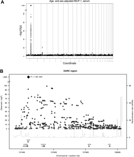
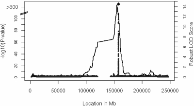
To identify the genetic basis of circulating concentrations of monocyte chemoattractant protein-1 (MCP-1), we conducted genome-wide association analyses for MCP-1 in 3 independent cohorts (n = 9598). The strongest association was for serum MCP-1 with a nonsynonymous polymorphism, rs12075 (Asp42Gly) in DARC, the gene for Duffy antigen receptor for chemokines, a known vascular reservoir of proinflammatory cytokines (minor allele frequency, 45.6%; P < 1.0 * 10(-323)). This association was supported by family-based genetic linkage at a locus encompassing the DARC gene (genome-wide P = 8.0 * 10(-13)). Asp42Gly accounted for approximately 20% of the variability in serum MCP-1 concentrations and also was associated with serum concentrations of interleukin-8 and RANTES. While exploring a lack of association between this polymorphism and EDTA plasma MCP-1 concentrations (P = .82), we determined that both clotting and exogenous heparan sulfate (unfractionated heparin) released substantial amounts of MCP-1 from Darc. Quantitative immunoflow cytometry failed to identify meaningful Asp42Gly-associated differences in Darc expression, suggesting that a functional change is responsible for the differential cytokine binding. We conclude that Asp42Gly is a major regulator of erythrocyte Darc-mediated cytokine binding and thereby the circulating concentrations of several proinflammatory cytokines. We have also identified for the first time 2 mechanisms for the release of reservoir chemokines with possible clinical implications.
Figures
Comment in
-
The DARC side of GWAS.Blood. 2010 Jul 1;115(26):5285-6. doi: 10.1182/blood-2010-02-263699. Blood. 2010. PMID: 20595521 No abstract available.
References
-
- Tracy RP. Thrombin, inflammation, and cardiovascular disease: an epidemiologic perspective. Chest. 2003;124(3 suppl):49S–57S. - PubMed
-
- Melgarejo E, Medina MA, Sanchez-Jimenez F, Urdiales JL. Monocyte chemoattractant protein-1: a key mediator in inflammatory processes. Int J Biochem Cell Biol. 2009;41(5):998–1001. - PubMed
-
- Neote K, Darbonne W, Ogez J, Horuk R, Schall TJ. Identification of a promiscuous inflammatory peptide receptor on the surface of red blood cells. J Biol Chem. 1993;268(17):12247–12249. - PubMed
Publication types
MeSH terms
Substances
Grants and funding
- UL1RR025005/RR/NCRR NIH HHS/United States
- R21 DA027021/DA/NIDA NIH HHS/United States
- R01 HL104156/HL/NHLBI NIH HHS/United States
- N01-HC-55022/HC/NHLBI NIH HHS/United States
- N01-HC-55016/HC/NHLBI NIH HHS/United States
- DK080739/DK/NIDDK NIH HHS/United States
- N01-HC-55021/HC/NHLBI NIH HHS/United States
- 2K24HL04334/HL/NHLBI NIH HHS/United States
- R01HL087641/HL/NHLBI NIH HHS/United States
- N01-HC-55015/HC/NHLBI NIH HHS/United States
- N01-HC-55020/HC/NHLBI NIH HHS/United States
- HL064753/HL/NHLBI NIH HHS/United States
- 1S10RR163736-01A1/RR/NCRR NIH HHS/United States
- R01 HL077449/HL/NHLBI NIH HHS/United States
- N01-HC-55018/HC/NHLBI NIH HHS/United States
- R01HL086694/HL/NHLBI NIH HHS/United States
- K24 HL105780/HL/NHLBI NIH HHS/United States
- R01HL59367/HL/NHLBI NIH HHS/United States
- AG028321/AG/NIA NIH HHS/United States
- U01HG004402/HG/NHGRI NIH HHS/United States
- HL093328/HL/NHLBI NIH HHS/United States
- N01-HC-55019/HC/NHLBI NIH HHS/United States
- 6R01-NS 17950/NS/NINDS NIH HHS/United States
- HL076784/HL/NHLBI NIH HHS/United States
LinkOut - more resources
Full Text Sources
Other Literature Sources
Research Materials
Miscellaneous
