. 2010 Mar;18(3):570-8.
doi: 10.1038/mt.2009.292. Epub 2009 Dec 29.

Directed evolution of a novel adeno-associated virus (AAV) vector that crosses the seizure-compromised blood-brain barrier (BBB)

Affiliations

Directed evolution of a novel adeno-associated virus (AAV) vector that crosses the seizure-compromised blood-brain barrier (BBB)

Steven J Gray et al. Mol Ther. 2010 Mar.

Erratum in

Abstract

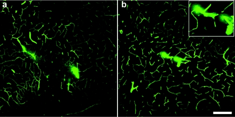
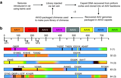
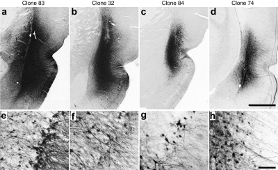
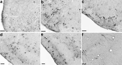
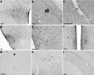
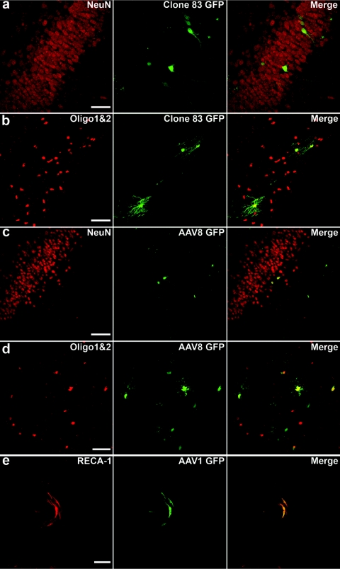
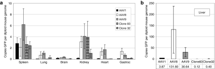
DNA shuffling and directed evolution were employed to develop a novel adeno-associated virus (AAV) vector capable of crossing the seizure-compromised blood-brain barrier (BBB) and transducing cells in the brain. Capsid DNA from AAV serotypes 1-6, 8, and 9 were shuffled and recombined to create a library of chimeric AAVs. One day after kainic acid-induced limbic seizure activity in rats, the virus library was infused intravenously (i.v.), and 3 days later, neuron-rich cells were mechanically dissociated from seizure-sensitive brain sites, collected and viral DNA extracted. After three cycles of selection, green fluorescent protein (GFP)-packaged clones were administered directly into brain or i.v. 1 day after kainic acid-induced seizures. Several clones that were effective after intracranial administration did not transduce brain cells after the i.v. administration. However, two clones (32 and 83) transduced the cells after direct brain infusion and after i.v. administration transduced the cells that were localized to the piriform cortex and ventral hippocampus, areas exhibiting a seizure-compromised BBB. No transduction occurred in areas devoid of BBB compromise. Only one parental serotype (AAV8) exhibited a similar expression profile, but the biodistribution of 32 and 83 diverged dramatically from this parental serotype. Thus, novel AAV vectors have been created that can selectively cross the seizure-compromised BBB and transduce cells.


Figures

<b>Figure 1</b>
Figure 1
Blood–brain barrier integrity is disrupted 24 hours after kainic acid administration and class IV seizures. Confocal micrographs of cerebral blood vessels perfused with fluorescein isothiocyanate–albumin illustrating irregular, cloud-like patches of albumin leakage in the (a) piriform cortex and (b) ventral hippocampus. Inset in b shows magnified image of microvascular compromise in the ventral hippocampus. Bar = 200 µm for a and b at equal magnification.
<b>Figure 2</b>
Figure 2
Directed evolution and recovery of chimeric clones. (a) Using a shuffled library based on adeno-associated virus (AAV) 1–6, 8, and 9, a directed evolution strategy was employed to select for an AAV capsid that can cross the seizure-compromised blood–brain barrier (BBB) and transduce cells in the areas of seizure influence. (b) Viable recovered clones that were tested in these studies. The scale at the top corresponds to the amino acid residues, with start sites for VP1, VP2, and VP3 indicated. A color-coding scheme is used to indicate the overall serotype composition at each location, and graded colors within each clone indicate regions of shared identity between neighboring serotypes. Where AAV1 and 6 are indistinguishable by amino acid sequence, a double stripe is drawn. Missense point mutations are shown as thin vertical lines, with the color corresponding to a serotype with that amino acid or a black line to indicate an amino acid not present in any parent. To the right of each amino acid map is the number assigned to that clone and the round from which it was recovered (in parentheses). wt, wild type.
<b>Figure 3</b>
Figure 3
Immunohistochemical analysis of green fluorescent protein transgene expression, 2 weeks following injections of infectious clones into piriform cortex. (a–d) Low-power photomicrographs of representative coronal sections showing the maximal dorsoventral extent of transduction around the site of injection. Bar = 1 mm for a–d at equal magnification. (e–h) Higher power images from sites depicted in a–d, demonstrating predominant neuronal tropism. Bar = 50 µm for e–h at equal magnification. (a,e) Clone 83; (b,f) clone 32; (c,g) clone 84; (d,h) clone 74.
<b>Figure 4</b>
Figure 4
Transduction patterns of clones 32 and 83 in rat brain two weeks after class IV seizures and intravenous vector administration. (a) Representative coronal section illustrating green fluorescent protein (GFP)–positive cells transduced by clone 32. Although not shown here, systemic injection of clone 32 results in GFP-positive cells throughout the rostro-caudal extent of the piriform cortex. (b–f) After intravenous injection of clone 83, GFP-positive cells were found in greater numbers compared to clone 32 throughout the full rostral–caudal extent of the piriform cortex as well as in the ventral hippocampus. (b) rostral piriform, 1.0 mm and (c) 0.2 mm anterior to bregma; (d) middle piriform, 2.2 mm posterior to bregma; (e) caudal piriform, 3.6 mm posterior to bregma; (f) and ventral hippocampus, 5.8 mm posterior to bregma. Bar = 100 µm for all panels.
<b>Figure 5</b>
Figure 5
Transduction patterns of adeno-associated virus (AAV)1, 8, and 9 in rat brain 2 weeks after class IV seizures and intravenous vector administration. (a–c) Systemic injection of AAV1 resulted in transduction of endothelial cells predominantly, with sporadic labeling of neurons and glia throughout the brain. (a) Neurons in the paraventricular nucleus of the hypothalamus; (b) green fluorescent protein (GFP)-labeled cells (presumably astrocytes) surrounding a blood vessel in the hippocampus; (c) endothelial cells lining a capillary in the motor cortex. (d–f) Intravenous injection of AAV8 resulted in a pattern of GFP labeling that was similar to, but more widespread than clones 32 and 83. (d) Middle piriform cortex, 2.2 mm posterior to bregma; (e) caudal piriform cortex, 3.2 mm posterior to bregma; (f) neuron-like cells in the medial preoptic nucleus of the hypothalamus. (g–i) Systemic injection of AAV9 resulted in scattered GFP-labeled cells throughout the brain. (g) Cells with glial and neuronal morphology in motor cortex; (h) a cluster of neurons in the nucleus of the horizontal limb of the diagonal band; (i) neurons in the dorsal hippocampus. Bar = 100 µm for all panels.
<b>Figure 6</b>
Figure 6
Clone 83 and adeno-associated virus (AAV)8 transduce neurons and oligodendrocytes, whereas AAV1 transduces primarily endothelial cells after intravenous administration and kainic acid injection. Fluorescence confocal micrographs illustrating clone 83-transduced cells [green fluorescent protein (GFP), green] colabel (a) with the neuronal marker NeuN in ventral hippocampus, and (b) with the oligodendrocyte markers Olig1 and Olig2 in the piriform cortex. Colabeling of GFP transduced by (c) AAV8 with NeuN, and (d) with Olig1 and Olig2, both in the piriform cortex. (e) The endothelial marker RECA-1 colocalizes with GFP-positive cells transduced by AAV1 in a large blood vessel near the cortical surface. Bar = 50 µm for all panels.
<b>Figure 7</b>
Figure 7
Biodistribution of clones 32 and 83. Mice were injected intravenously with 5 × 10¹⁰ vector genomes of each serotype, then after 10 days the mice were killed and DNA was isolated from the brain, heart, liver, spleen, lung, kidney, and gastrocnemius muscle. Values were calculated as copies of GFP per diploid copy of mouse genomic DNA (laminB2 locus). (a) Spleen, lung, brain, kidney, heart, and gastrocnemius muscle. (b) Biodistribution in liver (note difference in scale). Error bars indicate standard deviation. Numerical values for all groups and P values are provided in Supplementary Tables S2 and S3, respectively.

References

    1. Haberman RP, Samulski RJ., and , McCown TJ. Attenuation of seizures and neuronal death by adeno-associated virus vector galanin expression and secretion. Nat Med. 2003;9:1076–1080. - PubMed
    1. McCown TJ. Adeno-associated virus-mediated expression and constitutive secretion of galanin suppresses limbic seizure activity in vivo. Mol Ther. 2006;14:63–68. - PubMed
    1. Richichi R, Lin ED, Stefanin D, Colella D, Ravizza T, Grignaschi G, et al. Anticonvulsant and antiepileptic effects mediated by adeno-associated virus vector neuropeptide Y expression in the rat hippocampus. J Neurosci. 2004;24:3051–3059. - PMC - PubMed
    1. Foti SB, Russek ST, Brooks-Kayal AR., and , McCown TJ.2009Viral vector gene therapy for epilepsyIn: Baraban, SC (ed). Animal Models of Epilepsy: Methods and Innovations Humana Press: New York; 235–250.
    1. Maheshri N, Koerber JT, Kaspar BK., and , Schaffer DV. Directed evolution of adeno-associated virus yields enhanced gene delivery vectors. Nat Biotechnol. 2006;24:198–204. - PubMed

Publication types