

Expression of Semaphorin 3F and Its Receptors in Epithelial Ovarian Cancer, Fallopian Tubes, and Secondary Müllerian Tissues
- PMID: 20041133
- PMCID: PMC2796214
- DOI: 10.1155/2009/730739
Expression of Semaphorin 3F and Its Receptors in Epithelial Ovarian Cancer, Fallopian Tubes, and Secondary Müllerian Tissues
Abstract
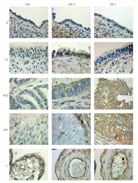
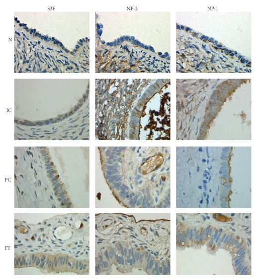
While semaphorins and their receptors appear to play a role in tumor carcinogenesis, little is known about the role of semaphorin 3F (S3F) in epithelial ovarian cancer (EOC) development. Therefore, we sought to determine the clinical relationship between S3F and its receptors, neuropilin-2 (NP-2) and neuropilin-1 (NP-1) with EOC progression. We analyzed the immunohistological expression of S3F, NP-2, and NP-1 in clinical specimens of normal ovaries (N), benign cystadenomas (Cy), well-differentiated adenocarcinomas (WD), poorly-differentiated adenocarcinomas (PD), inclusion cysts (IC), paraovarian cysts (PC), and fallopian tubes (FT). Tissue sections were evaluated for staining intensity and percentage of immunoreactive epithelia. We found that expression of S3F and NP-2 decreased while NP-1 expression increased with EOC progression. Interestingly, we also found elevated expression of S3F, NP-2, and NP-1 in epithelia of ICs, PCs, and FT. Our findings indicate that loss or deregulation of semaphorin signaling may play an important role in EOC development.
Figures


Similar articles
-
Semaphorin-3F is an inhibitor of tumor angiogenesis.Cancer Res. 2004 Feb 1;64(3):1008-15. doi: 10.1158/0008-5472.can-03-3090. Cancer Res. 2004. PMID: 14871832
-
Ontogeny of semaphorins 3A and 3F and their receptors neuropilins 1 and 2 in the kidney.Mech Dev. 2002 Dec;119 Suppl 1:S149-53. doi: 10.1016/s0925-4773(03)00108-4. Mech Dev. 2002. PMID: 14516677
-
Ovarian cancer and normal fallopian tube high WFDC2 expression does not correlate with HE4 serum level.Ginekol Pol. 2015 May;86(5):335-9. doi: 10.17772/gp/2418. Ginekol Pol. 2015. PMID: 26117969
-
Semaphorins in cancer.Front Biosci. 2005 Jan 1;10:751-60. doi: 10.2741/1569. Print 2005 Jan 1. Front Biosci. 2005. PMID: 15569615 Review.
-
Ovarian Cancer: The Fallopian Tube as the Site of Origin and Opportunities for Prevention.Front Oncol. 2016 May 2;6:108. doi: 10.3389/fonc.2016.00108. eCollection 2016. Front Oncol. 2016. PMID: 27200296 Free PMC article. Review.
Cited by
-
Semaphorins in angiogenesis and tumor progression.Cold Spring Harb Perspect Med. 2012 Jan;2(1):a006718. doi: 10.1101/cshperspect.a006718. Cold Spring Harb Perspect Med. 2012. PMID: 22315716 Free PMC article. Review.
-
Differences in the Expression Pattern of mRNA Protein SEMA3F in Endometrial Cancer in vitro under Cisplatin Treatment.Curr Pharm Biotechnol. 2020;21(11):1119-1128. doi: 10.2174/1389201021666200416102540. Curr Pharm Biotechnol. 2020. PMID: 32297576 Free PMC article.
-
Overexpression of semaphorin 3A promotes tumor progression and predicts poor prognosis in hepatocellular carcinoma after curative resection.Oncotarget. 2016 Aug 9;7(32):51733-51746. doi: 10.18632/oncotarget.10104. Oncotarget. 2016. PMID: 27351132 Free PMC article.
-
Role of Neuropilin-2-mediated signaling axis in cancer progression and therapy resistance.Cancer Metastasis Rev. 2022 Sep;41(3):771-787. doi: 10.1007/s10555-022-10048-0. Epub 2022 Jul 1. Cancer Metastasis Rev. 2022. PMID: 35776228 Free PMC article. Review.
-
Orchestrating Resilience: How Neuropilin-2 and Macrophages Contribute to Cardiothoracic Disease.J Clin Med. 2024 Mar 1;13(5):1446. doi: 10.3390/jcm13051446. J Clin Med. 2024. PMID: 38592275 Free PMC article. Review.
References
-
- Jemal A, Siegel R, Ward E, et al. Cancer statistics, 2009. CA: A Cancer Journal for Clinicians. 2009;59:225–249. - PubMed
-
- Williams TI, Toups KL, Saggese DA, Kalli KR, Cliby WA, Muddiman DC. Epithelial ovarian cancer: disease etiology, treatment, detection, and investigational gene, metabolite, and protein biomarkers. Journal of Proteome Research. 2007;6:2936–2962. - PubMed
-
- Piek JMJ, Kenemans P, Verheijen RHM. Intraperitoneal serous adenocarcinoma: a critical appraisal of three hypotheses on its cause. American Journal of Obstetrics and Gynecology. 2004;191(3):718–732. - PubMed
-
- Neufeld G, Kessler O. The semaphorins: versatile regulators of tumour progression and tumour angiogenesis. Nature Reviews Cancer. 2008;8(8):632–645. - PubMed
LinkOut - more resources
Full Text Sources
Miscellaneous
