. 2009 Dec 24;4(12):e8461.
doi: 10.1371/journal.pone.0008461.

A gene expression signature of invasive potential in metastatic melanoma cells

Affiliations

A gene expression signature of invasive potential in metastatic melanoma cells

Aaron R Jeffs et al. PLoS One. .

Abstract

Background: We are investigating the molecular basis of melanoma by defining genomic characteristics that correlate with tumour phenotype in a novel panel of metastatic melanoma cell lines. The aim of this study is to identify new prognostic markers and therapeutic targets that might aid clinical cancer diagnosis and management.

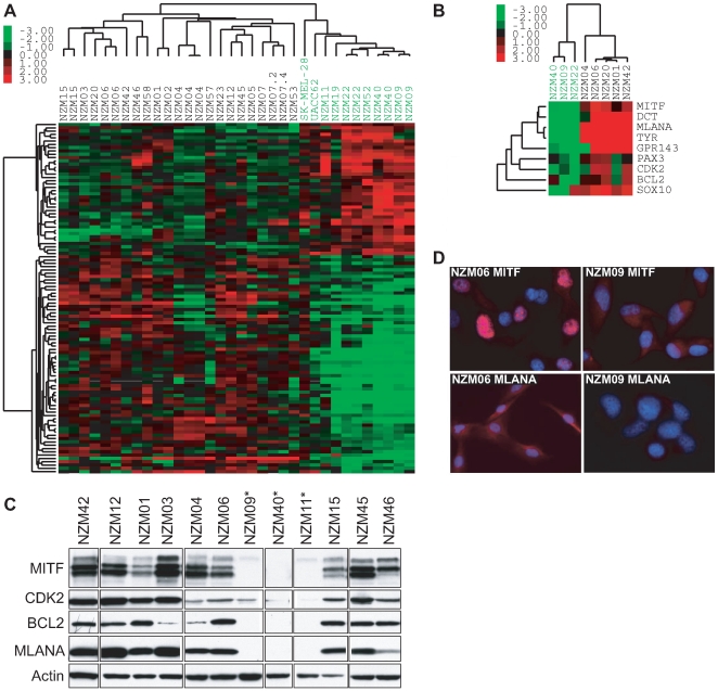
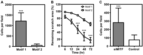
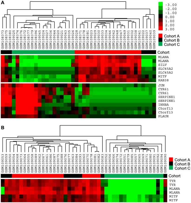
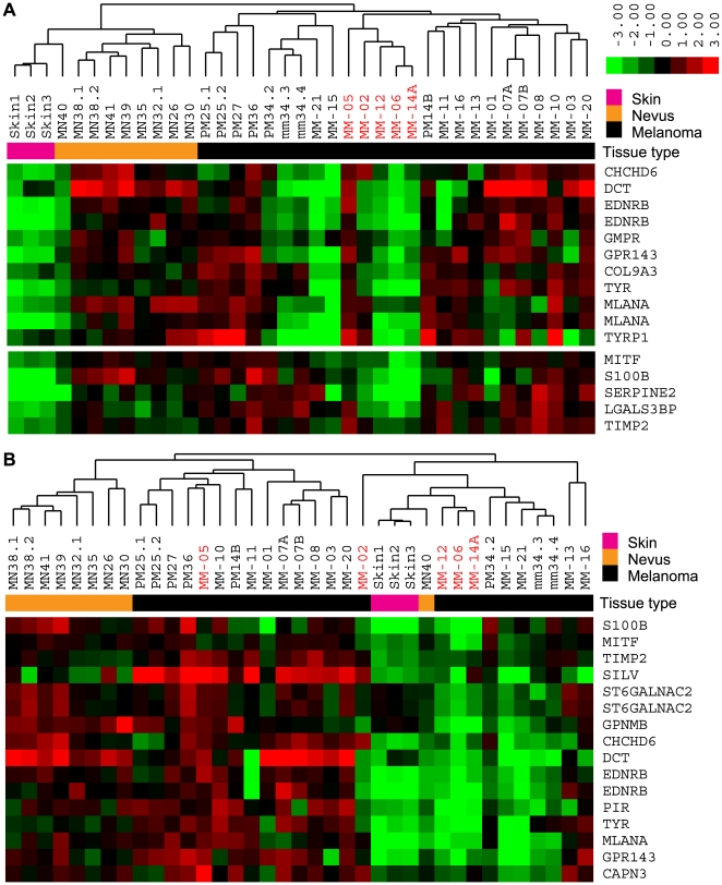
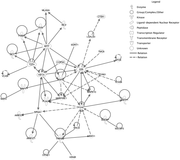
Principal findings: Global transcript profiling identified a signature featuring decreased expression of developmental and lineage specification genes including MITF, EDNRB, DCT, and TYR, and increased expression of genes involved in interaction with the extracellular environment, such as PLAUR, VCAN, and HIF1a. Migration assays showed that the gene signature correlated with the invasive potential of the cell lines, and external validation by using publicly available data indicated that tumours with the invasive gene signature were less melanocytic and may be more aggressive. The invasion signature could be detected in both primary and metastatic tumours suggesting that gene expression conferring increased invasive potential in melanoma may occur independently of tumour stage.

Conclusions: Our data supports the hypothesis that differential developmental gene expression may drive invasive potential in metastatic melanoma, and that melanoma heterogeneity may be explained by the differing capacity of melanoma cells to both withstand decreased expression of lineage specification genes and to respond to the tumour microenvironment. The invasion signature may provide new possibilities for predicting which primary tumours are more likely to metastasize, and which metastatic tumours might show a more aggressive clinical course.


Conflict of interest statement

Competing Interests: The authors have declared that no competing interests exist.

Figures

Figure 1
Figure 1. Expression profiling and validation.
(A) Unsupervised hierarchical clustering by using 96 genes identified by class comparison as differentially expressed between relatively higher or lower MITF-expressing melanoma cell lines classified the cell lines into two main groups (Motif 1, green label; Motif 2, black label). (B) qPCR validation of selected targets in a subset of Motif 1 and 2 NZM cell lines. Unsupervised clustering of qPCR data confirmed the Motif 1 (green) and 2 (black) classifications. (C) Protein expression of MITF targets CDK2, BCL2, and MLANA agreed with MITF transcript levels and array and qPCR cell line stratification. *Motif 1 cell lines. (D) Immunofluorescence showed stronger staining for MITF and MLANA in the Motif 2 NZM06 cells compared to Motif 1 NZM09 cells.
Figure 2
Figure 2. Motif 1 cell lines showed greater motility and migration in vitro.
(A) The number of Motif 1 cells (NZM09, NZM11, NZM22, NZM40, NZM52) that migrated through pored membranes in transwell (Boyden Chamber) assays was approximately 23-fold more than Motif 2 cell lines (NZM06, NZM12, NZM15, NZM42, NZM45; mean±SD is shown from the combined data of three separate experiments for each cell line; *** p<0.0001, t test). (B) Motif 1 cell lines NZM09 and NZM40 were significantly faster at wound repair in 2D scratch assays than Motif 2 NZM06 and NZM42 cell lines (mean±SEM; n = 3; *** p<0.001, two-way ANOVA). Representative movies of individual scratch assays for NZM09 and NZM42 are provided as supplementary Movie S1 and S2, respectively. (C) siRNA-mediated knockdown of MITF caused an almost 4-fold increase in migration of weakly invasive Motif 2 cell lines (NZM06, NZM15) in transwell assays compared to non-targeting siRNA controls (mean±SD from three separate experiments; *** p<0.0001, t test). MITF knockdown was confirmed by using qPCR and western blot (Figure S2).
Figure 3
Figure 3. External validation with independent cell line data.
Unsupervised clustering by using our 96 gene invasion signature on the combined Zurich and Philadelphia (A), or Mannheim (B) cell line data of Hoek et al. grouped cell lines into the cohorts of differing invasive potential originally identified by those authors, with Motif 1 corresponding to strongly invasive cohort C cell lines, and Motif 2 to weakly invasive cohort A cell lines.
Figure 4
Figure 4. External validation with independent tissue data.
(A) Unsupervised analysis by using our filtered gene list (572 transcripts) on the tissue data of Haqq et al. accurately classified skin, nevus, and melanoma tissue. MN, melanocytic nevus; PM, primary melanoma; MM, metastatic melanoma. (B) Application of our 96 gene invasion signature to the tissue data of Haqq et al. identified skin-like and nevus-like tumour samples representing Motif 1- and Motif 2-expressing tumour samples, respectively. Samples in red were derived from patients reported to have “type 1” tumours by the original authors.
Figure 5
Figure 5. A model linking expression of lineage specification and extracellular sensing genes to invasive potential in melanoma.
Pathway analysis suggested that melanoma invasive potential may be mediated by the intersection of MITF-driven transcriptional networks with pathways involved in HIF/JUN activation and response to hypoxia.

Similar articles

Cited by

References

    1. Bishop JN, Bataille V, Gavin A, Lens M, Marsden J, et al. The prevention, diagnosis, referral and management of melanoma of the skin: concise guidelines. Clin Med. 2007;7:283–290. - PMC - PubMed
    1. Hocker TL, Singh MK, Tsao H. Melanoma genetics and therapeutic approaches in the 21st century: moving from the benchside to the bedside. J Invest Dermatol. 2008;128:2575–2595. - PubMed
    1. Francken AB, Accortt NA, Shaw HM, Wiener M, Soong SJ, et al. Prognosis and determinants of outcome following locoregional or distant recurrence in patients with cutaneous melanoma. Ann Surg Oncol. 2008;15:1476–1484. - PubMed
    1. Jacobs LK, Lange JR, Balch CM. Sorting through the heterogeneity of recurrent melanoma. Ann Surg Oncol. 2008;15:1280–1281. - PMC - PubMed
    1. Balch CM, Soong SJ, Gershenwald JE, Thompson JF, Reintgen DS, et al. Prognostic factors analysis of 17,600 melanoma patients: validation of the American Joint Committee on Cancer melanoma staging system. J Clin Oncol. 2001;19:3622–3634. - PubMed

Publication types

MeSH terms