. 2009 Dec;5(12):e1000705.
doi: 10.1371/journal.ppat.1000705. Epub 2009 Dec 24.

A unique role for the host ESCRT proteins in replication of Tomato bushy stunt virus

Affiliations

A unique role for the host ESCRT proteins in replication of Tomato bushy stunt virus

Daniel Barajas et al. PLoS Pathog. 2009 Dec.

Abstract

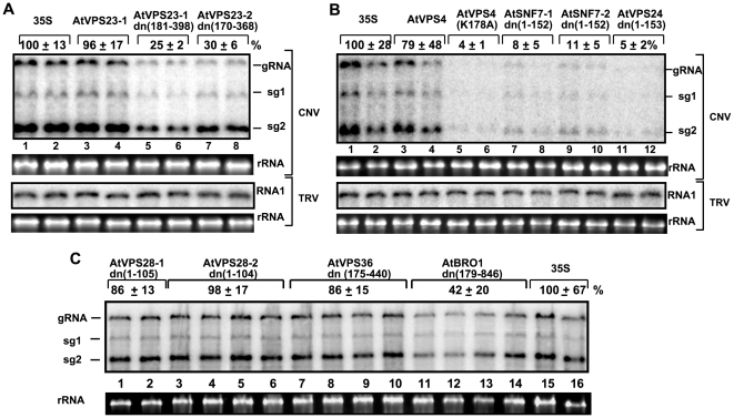
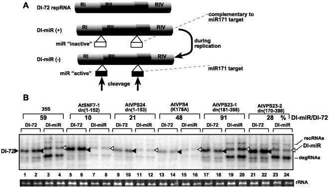
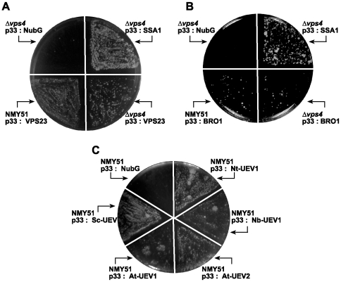
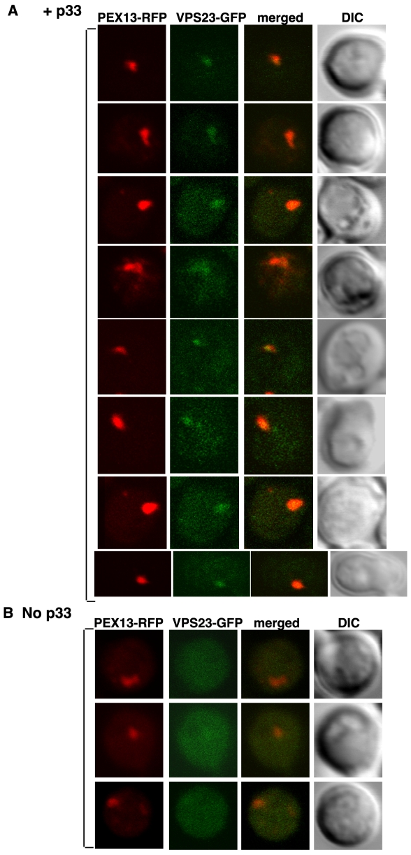
Plus-stranded RNA viruses replicate in infected cells by assembling viral replicase complexes consisting of viral- and host-coded proteins. Previous genome-wide screens with Tomato bushy stunt tombusvirus (TBSV) in a yeast model host revealed the involvement of seven ESCRT (endosomal sorting complexes required for transport) proteins in viral replication. In this paper, we show that the expression of dominant negative Vps23p, Vps24p, Snf7p, and Vps4p ESCRT factors inhibited virus replication in the plant host, suggesting that tombusviruses co-opt selected ESCRT proteins for the assembly of the viral replicase complex. We also show that TBSV p33 replication protein interacts with Vps23p ESCRT-I and Bro1p accessory ESCRT factors. The interaction with p33 leads to the recruitment of Vps23p to the peroxisomes, the sites of TBSV replication. The viral replicase showed reduced activity and the minus-stranded viral RNA in the replicase became more accessible to ribonuclease when derived from vps23Delta or vps24Delta yeast, suggesting that the protection of the viral RNA is compromised within the replicase complex assembled in the absence of ESCRT proteins. The recruitment of ESCRT proteins is needed for the precise assembly of the replicase complex, which might help the virus evade recognition by the host defense surveillance system and/or prevent viral RNA destruction by the gene silencing machinery.


Conflict of interest statement

The authors have declared that no competing interests exist.

Figures

Figure 1
Figure 1. Inhibition of tombusvirus RNA accumulation in plants by expression of dominant negative ESCRT mutants.
(A) Expression of N-terminal deletion mutants of the two homologous AtVps23p proteins (lanes 5–8) was done in N. benthamiana leaves, which were co-infiltrated with Agrobacterium carrying a plasmid to launch CNV replication from the 35S promoter. The control samples were obtained from leaves expressing no proteins (35S, lanes 1-2) or the full-length AtVps23p (lanes 3–4). Total RNA was extracted from leaves 2.5 days after agroinfiltration. The accumulation of CNV gRNA and subgenomic (sg)RNAs in N. benthamiana leaves was measured by Northern blotting (Top panel). The ribosomal RNA (rRNA) was used as a loading control and shown in agarose gel stained with ethidium-bromide (Second panel). The bottom two panels show the lack of inhibition of TRV RNA1 accumulation by the expression of the above proteins. TRV infection was launched by agroinfiltration as described above for CNV. (B) Blocking tombusvirus RNA replication in plants by expression of the dominant negative mutant of AtVps4p [named AtVps4p(K178A)] (lanes 5–6), and C-terminal deletion mutants of two homologous AtSnf7p and one AtVps24p ESCRT-III proteins. See further details in panel A. (C) The expression of the C-terminal deletion mutants of two homologous AtVps28p and the N-terminal deletion mutants of AtVps36p and AtBro1p ESCRT proteins in plants was done from the constitutive 35S promoter. See further details in panel A.
Figure 2
Figure 2. The isolated tombusvirus replicase preparations from N. benthamiana plants expressing dominant negative ESCRT factors show low activity in vitro.
Top panel: Denaturing PAGE of in vitro replicase activity in the membrane-enriched fraction from co-infiltrated leaves expressing p33, p92pol, DI-72 repRNA, p19 (suppressor of gene silencing) and the shown dominant negative ESCRT factors using the co-purified repRNA template. The lack of replicase activity in the absence of p33 (lane 11) or p92pol (lane 12) demonstrates that the in vitro replication is tombusvirus specific. Bottom panel: Western blot analysis of p33 and p92pol levels in the above membrane-enriched fractions was performed with anti-p33 antibody.
Figure 3
Figure 3. Reduced activity of the tombusvirus replicase assembled in yeast with deletion of selected ESCRT genes.
(A) Denaturing PAGE analysis of in vitro replicase activity in the membrane-enriched fraction from wt and vps23Δ yeast using the co-purified repRNA. Note that this image shows the repRNAs made by the replicase in vitro. Asterisk marks a recombinant RNA species formed. Bottom panel shows a Western blot of p33 in the replicase preparations as seen in the top panel. (B) Decreased replication of TBSV repRNA in yeast extracts prepared from wt, vps4Δ, snf7Δ or vps24Δ yeast strains expressing p33 and p92pol. The yeast extracts were programmed with DI-72(+)repRNA and the radiolabeled in vitro repRNA products were detected via denaturing PAGE analysis. (C) Increased RNase sensitivity of TBSV minus-strand (−)repRNA during replication in a membrane-enriched replicase preparation obtained from WT, vps23Δ or vps24Δ yeast. At the end of the assays, the replicase preparations were treated with RNase A for 5 min, followed by inactivation with phenol-chloroform. The (−)repRNA protected in the replicase complex from the ribonuclease was detected using a 32P-UTP labeled probe. The untreated preparation was chosen as 100%. Note that the (−)repRNA is protected from RNase degradation by the membrane-associated viral replicase complex.
Figure 4
Figure 4. Increased sensitivity of repRNA replication to targeted degradation by RNAi in plants expressing dominant negative ESCRT-III mutants.
(A) A schematic representation of constructs used as repRNAs. Two copies of the 21 nt long miR171 target sequence were inserted into DI-72 repRNA to generate DI-miR repRNA as shown. Note that both copies of the miR171 target sequence were present in the (−)strand RNA generated during repRNA replication only in plants agroinfiltrated with constructs expressing p33/p92/dominant negative ESCRT proteins and DI-72 or DI-miR repRNAs. (B) Northern blot analysis shows reduced accumulation of DI-miR repRNA in N. benthamiana plants expressing dominant negative ESCRT-III or Vps4p mutants. DI-72 repRNA is depicted with a black arrowhead, while DI-miR repRNA is marked with an open arrowhead. The percentage of DI-miR repRNA accumulation was calculated based on DI-72 repRNA levels (taken as 100% for each set). Note that the low level DI-miR repRNA accumulation suggests that the RNAi machinery destroyed most of the (−)repRNA present in the replicase complex. recRNA represents recombinant repRNAs, while degRNA is derived from partially degraded repRNA. Note that degRNA can replicate in plants, so it does not represent the original cleaved repRNA.
Figure 5
Figure 5. Interaction between p33 replication protein and Vps23p ESCRT-I and Bro1p accessory ESCRT proteins.
The split ubiquitin assay was used to test binding between p33 and (A) Vps23p; (B) Bro1p, or (C) the N-terminal UEV domain of the yeast Sc-Vps23p or two Arabidopsis and two Nicotiana homologs in wt (NMY51) or vps4Δ yeast. The bait p33 was co-expressed with the shown prey proteins. SSA1 (HSP70 chaperone), and the empty prey vector (NubG) were used as positive and negative controls, respectively.
Figure 6
Figure 6. Co-purification of the p33 replication protein with the UEV domain of Vps23p ESCRT-I or Bro1p accessory ESCRT proteins.
Top panels: Western blot analysis of co-purified p33 and either (A) the UEV domain or (B) Bro1p protein. The FLAG/6xHis-tagged p33HF was purified from yeast extracts using a FLAG-affinity column. UEV and Bro1p, both tagged with 6xHA, were detected with anti-HA antibody. Middle panels: Western blot of purified p33HF detected with anti-FLAG antibody. Bottom panels: Western blot of HA-tagged UEV and Bro1p in the total yeast extract using anti-HA antibody.
Figure 7
Figure 7. Partial re-distribution of Vps23p to the yeast peroxisomal membranes in the presence of p33.
(A) Confocal laser microscopy images show the subcellular localization of Vps23p-GFP in the presence of p33 expressed from CUP1 promoter for 15–45 minutes in yeast strain DKY79 (VPS23:GFP, vps4Δ; vps27Δ). The peroxisomes were visualized with Pex13p-RFP marker. The merged images show the co-localization of Vps23p-GFP and Pex13p-RFP marker. DIC (differential interference contrast) images are shown on the right. Each row represents a separate yeast cell. (B) Cytosolic localization of Vps23p-GFP in the absence of p33. Yeast was grown under similar conditions and images were taken as in panel A.
Figure 8
Figure 8. A model for the role ESCRT proteins in tombusvirus replication.
Recruitment of Vps23p and/or Bro1p to the peroxisomal membrane by a small fraction of p33 is suggested to lead to the recruitment of additional ESCRT factors. Then, ESCRT-III and Vps4p factors facilitate the precise assembly of the replicase that is needed to prevent the recognition of the virus by the host surveillance system and prevent the destruction of the viral RNAs in the spherule by the host gene silencing machinery.

Similar articles

Cited by

References

    1. Nagy PD. Yeast as a model host to explore plant virus-host interactions. Annu Rev Phytopathol. 2008;46:217–242. - PubMed
    1. Ahlquist P, Noueiry AO, Lee WM, Kushner DB, Dye BT. Host factors in positive-strand RNA virus genome replication. J Virol. 2003;77:8181–8186. - PMC - PubMed
    1. Noueiry AO, Ahlquist P. Brome Mosaic Virus RNA Replication: Revealing the Role of the Host in RNA Virus Replication. Annu Rev Phytopathol 2003 - PubMed
    1. Nagy PD, Pogany J. Multiple roles of viral replication proteins in plant RNA virus replication. Methods Mol Biol. 2008;451:55–68. - PubMed
    1. Panavas T, Serviene E, Brasher J, Nagy PD. Yeast genome-wide screen reveals dissimilar sets of host genes affecting replication of RNA viruses. Proc Natl Acad Sci U S A. 2005;102:7326–7331. - PMC - PubMed

Publication types

MeSH terms

Substances

LinkOut - more resources