. 2009 Dec 29;4(12):e8484.
doi: 10.1371/journal.pone.0008484.

Fiber mediated receptor masking in non-infected bystander cells restricts adenovirus cell killing effect but promotes adenovirus host co-existence

Affiliations

Fiber mediated receptor masking in non-infected bystander cells restricts adenovirus cell killing effect but promotes adenovirus host co-existence

Johan Rebetz et al. PLoS One. .

Abstract

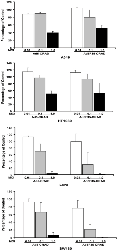
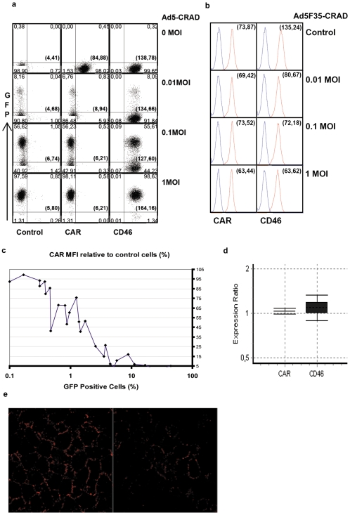
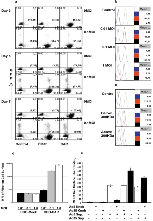
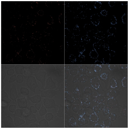
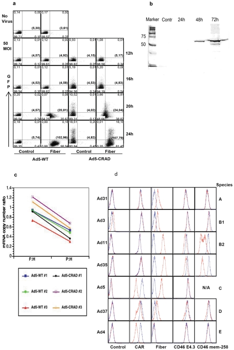
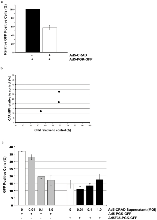
The basic concept of conditionally replicating adenoviruses (CRAD) as oncolytic agents is that progenies generated from each round of infection will disperse, infect and kill new cancer cells. However, CRAD has only inhibited, but not eradicated tumor growth in xenograft tumor therapy, and CRAD therapy has had only marginal clinical benefit to cancer patients. Here, we found that CRAD propagation and cancer cell survival co-existed for long periods of time when infection was initiated at low multiplicity of infection (MOI), and cancer cell killing was inefficient and slow compared to the assumed cell killing effect upon infection at high MOI. Excessive production of fiber molecules from initial CRAD infection of only 1 to 2% cancer cells and their release prior to the viral particle itself caused a tropism-specific receptor masking in both infected and non-infected bystander cells. Consequently, the non-infected bystander cells were inefficiently bound and infected by CRAD progenies. Further, fiber overproduction with concomitant restriction of adenovirus spread was observed in xenograft cancer therapy models. Besides the CAR-binding Ad4, Ad5, and Ad37, infection with CD46-binding Ad35 and Ad11 also caused receptor masking. Fiber overproduction and its resulting receptor masking thus play a key role in limiting CRAD functionality, but potentially promote adenovirus and host cell co-existence. These findings also give important clues for understanding mechanisms underlying the natural infection course of various adenoviruses.


Conflict of interest statement

Competing Interests: The authors have declared that no competing interests exist.

Figures

Figure 1
Figure 1. Cancer cell killing of CRAD infection at low MOI.
Mean ± SD (n = 3) of total cell numbers of CRAD infected cell cultures relative to non-infected control cultures at one-week post infection are shown.
Figure 2
Figure 2. Decrease of receptor detectibility following CRAD replication in a small fraction of cells.
(A) GFP expression and CAR, CD46 intensity in A549 cells at one week following Ad5-CRAD infection at indicated MOI. Numbers in each quadrate represent the phenotypic distribution of the cells according to GFP expression and CD46 or CAR mAb staining intensity. The numbers in brackets represent the mean fluorescence intensity (MFI) of CAR or CD46 staining of all cells. Data are representative for (C). (B) CAR and CD46 intensity in A549 cells at one week following Ad5F35-CRAD infection. The numbers in brackets represent the MFI of CAR or CD46 staining (red line in each histogram) of all cells. The blue lines represent isostype control antibody staining. (C) Inverse correlation between CAR intensity and the percentages of GFP+ cells following Ad5-CRAD infection at low MOIs. Each dot represents one independent assay for GFP expression and CAR MFI relative to the control non-infected cells. (D) The ratios of CAR or CD46 mRNA expression level (filled box, mean ± SD) and its 95% confidence intervals between Ad5-CRAD infected (with ∼2% GFP+ cells) and non-infected A549 cultures. (E) Representative CLSM analyses of RcmB binding in A549 control culture (left panel) or cultures at day 6 post infection with Ad5-CRAD at a MOI of 1.0 with ∼1.5% cells being GFP+ (right panel).
Figure 3
Figure 3. Fiber binding and receptor masking in infected and non-infected cells.
(A) Progressive fiber binding and concomitant decrease in CAR intensity in both infected and non-infected A549 cells were similar following infection with Ad5-CRAD at low MOI. Representative data from 3 independent studies are shown. (B) Supernatants from Ad5-CRAD infected A549 cultures conferred cell surface fiber binding and concomitant decrease in CAR intensity in non-infected A549 cells. Non-infected cells were incubated for 2 hr with A549 culture supernatants harvested at one week post infection with Ad5-CRAD at the indicated MOI, washed and analyzed for CAR and fiber intensity. Data shown are representative histograms of 3 experiments performed at 37°C. The MFIs of isotype control (blue lines), CAR (red lines) and fiber (black lines) staining of all cells are indicated at the right part of each histogram. (C) Free fiber molecules in <300 KDa supernatant fraction from Ad5-CRAD infected A549 cultures conferred the same effect as in (B). Supernatants used in (B) were centrifuged at 108 000 g for 1 hr, fractioned through a membrane with 300 KDa cut-off, and subsequently used in binding experiments as in (B). (D) Supernatants from A549 cell cultures at one week following infection with Ad5-CRAD at the indicated MOIs conferred fiber binding only to CAR expressing CHO cells. The mean ± SD (n = 2) of fiber binding MFI are shown. (E) Supernatant fiber binding to fresh A549 cells was inhibited by recombinant Ad5 or Ad35 fiber knob molecules in a tropism specific manner. The mean ± SD (n = 2) of fiber binding MFI to A549 cells are shown.
Figure 4
Figure 4. CLSM analysis of cell surface CAR expression and fiber binding.
A549 cultures at 6 days post infection with Ad5-CRAD at a MOI of 1.0 were co-stained for cell surface CAR expression with the polyclonal rabbit anti-CAR 72 antibody (upper left) and fiber binding with mAb 4D2 (upper right) and analyzed in CLSM. The merged images (lower right) demonstrate the co-localization (white) of CAR and fiber molecules on cell surface.
Figure 5
Figure 5. Mechanisms of fiber production during adenovirus life cycle.
(A) Fiber secretion prior to adenoviral particle release. A549 cells were infected with WT Ad5 or Ad5-CRAD at a MOI of 50, cell surface fiber binding and GFP expression were assessed at the indicated time points post infection. Data are representative for A549 cell infection experiments. (B) Release of fiber upon cell lysis. A549 cells were infected with WT Ad5 at a MOI of 10 in DMEM containing 2% FCS. Forty µg of supernatant proteins harvested from the control non-infected culture, or from infected cultures at 24, 48 and 72 hr post infection were analyzed in Western-blot with 4D2 anti fiber mAb. (C) Abundance of fiber mRNA in Ad5 life cycle. Total RNA was extracted from A549 cells at 20 hr post infection with WT Ad5 or Ad5-CRAD as in (A), and analyzed for fiber, penton and hexon mRNA copy numbers in real-time RT-PCR. The paired ratios of fiber to hexon (F:H) and penton to hexon (P:H) mRNA copy numbers from each infection are depicted. (D) Fiber overproduction and receptor masking of various adenovirus serotypes. A549 cells were infected with indicated WT adenoviruses at 500 (Ad31, Ad3, and Ad37), 50 (Ad4), or 10 (Ad11, Ad35 and Ad5) viral particles per cell. Cell surface fiber binding and receptor intensity were analyzed upon visible CPE or day 10 post infection. In each histogram, the red and blue lines represent staining with infected and non-infected control cultures, respectively.
Figure 6
Figure 6. Consequences of fiber mediated receptor masking for CRAD infection.
(A) Infectivity of receptor masked A549 cells by adenoviral vectors. A549 culture was first infected with Ad5-CRAD at a MOI 1.0. Six days later, cells were further infected with Ad5-PGK-GFP at a MOI of 10 and GFP expression was assessed 24 hr later. Data shown are the relative mean ± SD (n = 3) of the percentages of GFP+ cells in cultures infected by both Ad5-CRAD and Ad5-PGK-GFP compared to control cultures infected by Ad5-PGK-GFP only. (B) Adenovirus binding capacity to receptor masked cells. CAR intensity and 35S-Ad5 binding capacity to cells of A549 culture at day 6 post Ad5-CRAD infection (MOI  = 1.0) relative to cells of non-infected control culture of three independent experiments are shown. (C) Supernatants from Ad5-CRAD infected A549 cells conferred tropism-specific low infectivity to fresh A549 cells. A549 cell cultures were first incubated with supernatants of Ad5-CRAD infected cultures as in Figure 3B. Following extensive washing, cells were further infected with Ad5-PGK-GFP or Ad5F35-PGK-GFP at a MOI of 10. GFP expression was measured 24 hr later. The mean ± SD (n = 3) of the percentages of GFP+ cells are shown. The percentages of GFP+ cells in cultures incubated with the indicated Ad5-CRAD infection supernatant alone were deducted from the values presented in the grey or black bars.
Figure 7
Figure 7. Extensive fiber production during CRAD treatment of xenograft tumors.
Following a single intratumoral injection of 1×10⁹ PFU CNHK500, A549 cell xenograft tumors were dissected at day 3, 7 and 10 post injection and consecutive sections were stained for fiber and hexon proteins. Representative staining of fiber and hexon proteins are shown (×200).
Figure 8
Figure 8. Effects of fiber overproduction and its resulting masking in CRAD cancer therapy and adenovirus propagation.
The initially infected cells secrete large amounts of fiber molecules prior to progeny virus release. Consequently, the receptors on bystander cells are masked by fiber molecules (red). Receptor masked cells cannot be efficiently infected by progeny viruses (blue) when these are released upon cell lysis. This mechanism inhibits CRAD cell killing effects, but promotes a prolonged adenovirus host co-existence.

Similar articles

Cited by

References

    1. Fox JP, Hall CE, Cooney MK. The Seattle Virus Watch. VII. Observations of adenovirus infections. Am J Epidemiol. 1977;105(4):362–386. - PubMed
    1. Garnett CT, Talekar G, Mahr JA, Huang W, Zhang Y, et al. Latent species C adenoviruses in human tonsil tissues. J Virol. 2009;83(6):2417–2428. - PMC - PubMed
    1. Heise C, Kirn DH. Replication-selective adenoviruses as oncolytic agents. J Clin Invest. 2000;105(7):847–851. - PMC - PubMed
    1. Lichtenstein DL, Wold WS. Experimental infections of humans with wild-type adenoviruses and with replication-competent adenovirus vectors: replication, safety, and transmission. Cancer Gene Ther. 2004;11(12):819–829. - PubMed
    1. Edqvist A, Rebetz J, Jaras M, Rydelius A, Skagerberg G, et al. Detection of cell cycle- and differentiation stage-dependent human telomerase reverse transcriptase expression in single living cancer cells. Mol Ther. 2006;14(1):139–148. - PubMed

Publication types

LinkOut - more resources