

Rational mutational analysis of a multidrug MFS transporter CaMdr1p of Candida albicans by employing a membrane environment based computational approach
- PMID: 20041202
- PMCID: PMC2789324
- DOI: 10.1371/journal.pcbi.1000624
Rational mutational analysis of a multidrug MFS transporter CaMdr1p of Candida albicans by employing a membrane environment based computational approach
Abstract
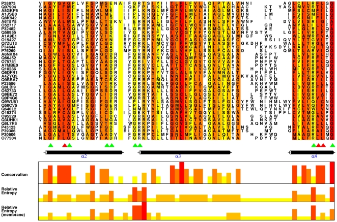
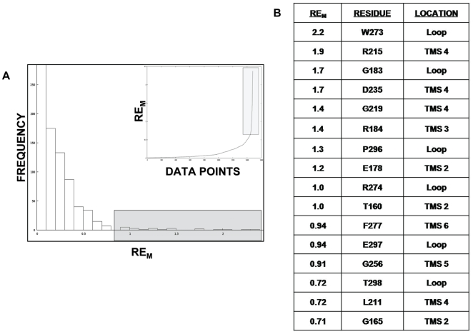
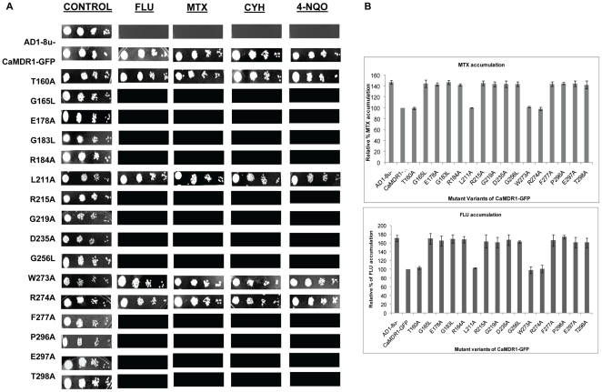
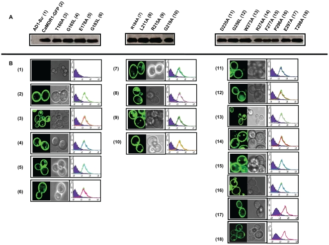
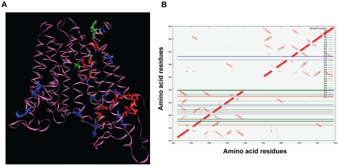
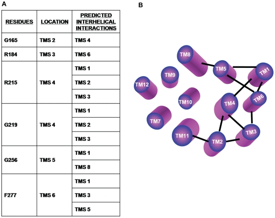
CaMdr1p is a multidrug MFS transporter of pathogenic Candida albicans. An over-expression of the gene encoding this protein is linked to clinically encountered azole resistance. In-depth knowledge of the structure and function of CaMdr1p is necessary for an effective design of modulators or inhibitors of this efflux transporter. Towards this goal, in this study, we have employed a membrane environment based computational approach to predict the functionally critical residues of CaMdr1p. For this, information theoretic scores which are variants of Relative Entropy (Modified Relative Entropy RE(M)) were calculated from Multiple Sequence Alignment (MSA) by separately considering distinct physico-chemical properties of transmembrane (TM) and inter-TM regions. The residues of CaMdr1p with high RE(M) which were predicted to be significantly important were subjected to site-directed mutational analysis. Interestingly, heterologous host Saccharomyces cerevisiae, over-expressing these mutant variants of CaMdr1p wherein these high RE(M) residues were replaced by either alanine or leucine, demonstrated increased susceptibility to tested drugs. The hypersensitivity to drugs was supported by abrogated substrate efflux mediated by mutant variant proteins and was not attributed to their poor expression or surface localization. Additionally, by employing a distance plot from a 3D deduced model of CaMdr1p, we could also predict the role of these functionally critical residues in maintaining apparent inter-helical interactions to provide the desired fold for the proper functioning of CaMdr1p. Residues predicted to be critical for function across the family were also found to be vital from other previously published studies, implying its wider application to other membrane protein families.
Conflict of interest statement
The authors have declared that no competing interests exist.
Figures
Similar articles
-
Employing information theoretic measures and mutagenesis to identify residues critical for drug-proton antiport function in Mdr1p of Candida albicans.PLoS One. 2010 Jun 10;5(6):e11041. doi: 10.1371/journal.pone.0011041. PLoS One. 2010. PMID: 20548793 Free PMC article.
-
Structure and function analysis of CaMdr1p, a major facilitator superfamily antifungal efflux transporter protein of Candida albicans: identification of amino acid residues critical for drug/H+ transport.Eukaryot Cell. 2007 Mar;6(3):443-53. doi: 10.1128/EC.00315-06. Epub 2007 Jan 5. Eukaryot Cell. 2007. PMID: 17209122 Free PMC article.
-
Specificity of drug transport mediated by CaMDR1: a major facilitator of Candida albicans.J Biosci. 2001 Sep;26(3):333-9. doi: 10.1007/BF02703742. J Biosci. 2001. PMID: 11568478
-
Candida Efflux ATPases and Antiporters in Clinical Drug Resistance.Adv Exp Med Biol. 2016;892:351-376. doi: 10.1007/978-3-319-25304-6_15. Adv Exp Med Biol. 2016. PMID: 26721282 Review.
-
The ABCs of Candida albicans Multidrug Transporter Cdr1.Eukaryot Cell. 2015 Dec;14(12):1154-64. doi: 10.1128/EC.00137-15. Epub 2015 Sep 25. Eukaryot Cell. 2015. PMID: 26407965 Free PMC article. Review.
Cited by
-
Employing information theoretic measures and mutagenesis to identify residues critical for drug-proton antiport function in Mdr1p of Candida albicans.PLoS One. 2010 Jun 10;5(6):e11041. doi: 10.1371/journal.pone.0011041. PLoS One. 2010. PMID: 20548793 Free PMC article.
-
Bacillus cereus efflux protein BC3310 - a multidrug transporter of the unknown major facilitator family, UMF-2.Front Microbiol. 2015 Oct 12;6:1063. doi: 10.3389/fmicb.2015.01063. eCollection 2015. Front Microbiol. 2015. PMID: 26528249 Free PMC article.
-
Host-induced gene silencing of a regulator of G protein signalling gene (VdRGS1) confers resistance to Verticillium wilt in cotton.Plant Biotechnol J. 2018 Feb 12;16(9):1629-43. doi: 10.1111/pbi.12900. Online ahead of print. Plant Biotechnol J. 2018. PMID: 29431919 Free PMC article.
-
Energy coupling mechanisms of MFS transporters.Protein Sci. 2015 Oct;24(10):1560-79. doi: 10.1002/pro.2759. Epub 2015 Sep 18. Protein Sci. 2015. PMID: 26234418 Free PMC article. Review.
-
Directed Mutational Strategies Reveal Drug Binding and Transport by the MDR Transporters of Candida albicans.J Fungi (Basel). 2021 Jan 20;7(2):68. doi: 10.3390/jof7020068. J Fungi (Basel). 2021. PMID: 33498218 Free PMC article. Review.
References
-
- Prasad R, Kapoor K. Multidrug resistance in yeast Candida. Int Rev Cytol. 2005;242:215–248. - PubMed
-
- Gaur M, Choudhury D, Prasad R. Complete inventory of ABC proteins in human pathogenic yeast, Candida albicans. J Mol Microbiol Biotechnol. 2005;9:3–15. - PubMed
-
- Smriti, Krishnamurthy S, Dixit BL, Gupta CM, Milewski S, et al. ABC transporters Cdr1p, Cdr2p and Cdr3p of a human pathogen Candida albicans are general phospholipid translocators. Yeast. 2002;19:303–318. - PubMed
Publication types
MeSH terms
Substances
LinkOut - more resources
Full Text Sources
Molecular Biology Databases
