. 2009 Dec;5(12):e1000704.
doi: 10.1371/journal.ppat.1000704. Epub 2009 Dec 24.

Molecular mimicry by an F-box effector of Legionella pneumophila hijacks a conserved polyubiquitination machinery within macrophages and protozoa

Affiliations

Molecular mimicry by an F-box effector of Legionella pneumophila hijacks a conserved polyubiquitination machinery within macrophages and protozoa

Christopher T Price et al. PLoS Pathog. 2009 Dec.

Abstract

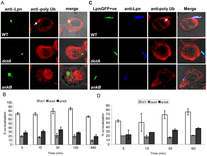
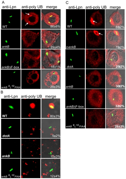
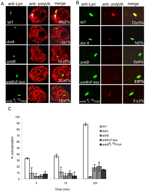
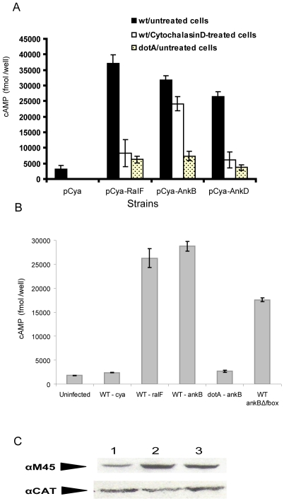
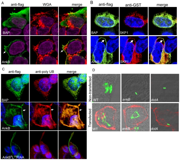
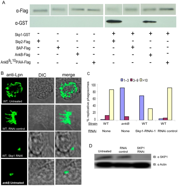
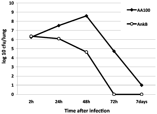
The ability of Legionella pneumophila to proliferate within various protozoa in the aquatic environment and in macrophages indicates a remarkable evolution and microbial exploitation of evolutionarily conserved eukaryotic processes. Ankyrin B (AnkB) of L. pneumophila is a non-canonical F-box-containing protein, and is the only known Dot/Icm-translocated effector of L. pneumophila essential for intra-vacuolar proliferation within both macrophages and protozoan hosts. We show that the F-box domain of AnkB and the (9)L(10)P conserved residues are essential for intracellular bacterial proliferation and for rapid acquisition of polyubiquitinated proteins by the Legionella-containing vacuole (LCV) within macrophages, Dictyostelium discoideum, and Acanthamoeba. Interestingly, translocation of AnkB and recruitment of polyubiquitinated proteins in macrophages and Acanthamoeba is rapidly triggered by extracellular bacteria within 5 min of bacterial attachment. Ectopically expressed AnkB within mammalian cells is localized to the periphery of the cell where it co-localizes with host SKP1 and recruits polyubiquitinated proteins, which results in restoration of intracellular growth to the ankB mutant similar to the parental strain. While an ectopically expressed AnkB-(9)L(10)P/AA variant is localized to the cell periphery, it does not recruit polyubiquitinated proteins and fails to trans-rescue the ankB mutant intracellular growth defect. Direct in vivo interaction of AnkB but not the AnkB-(9)L(10)P/AA variant with the host SKP1 is demonstrated. Importantly, RNAi-mediated silencing of expression of SKP1 renders the cells non-permissive for intracellular proliferation of L. pneumophila. The role of AnkB in exploitation of the polyubiquitination machinery is essential for intrapulmonary bacterial proliferation in the mouse model of Legionnaires' disease. Therefore, AnkB exhibits a novel molecular and functional mimicry of eukaryotic F-box proteins that exploits conserved polyubiquitination machinery for intracellular proliferation within evolutionarily distant hosts.


Conflict of interest statement

The authors have declared that no competing interests exist.

Figures

Figure 1
Figure 1. AnkB is essential for recruitment of polyubiquitinated proteins by the LCV and bacterial attachment to the macrophage plasma membrane is sufficient to trigger the recruitment process.
U937 cells were infected with the wild type (WT) L. pneumophila (Lpn) and the isogenic dotA or ankB mutants. A) Representative confocal microscopy images of infected U937 cells at 2 h post-infection for co-localization of the LCVs with polyubiquitinated (PolyUb) proteins. The arrow indicates heavy co-localization of polyubiquitin with the WT strain. B) Quantitation of the co-localization during infection at different time points. In panels C and D, the U937 cells were treated with 1 µM cytochalasin D for 30 min prior to infection, to inhibit bacterial entry. The cells were infected with GFP-expressing bacteria for 5, 15, 30 and 60 min. Prior to permeabilization, extracellular L. pneumophila were labeled with an anti-Lpn antibody (blue) resulting in a dual labeling of the extracellular bacteria (green and blue). Host polyubiquitin was labeled after permeabilization (red). C) Representative confocal microscopy images that show an extracellular WT L. pneumophila bacterium co-localized with high levels of polyubiquitinated proteins (arrow) at the site of bacterial attachment after 5 min of infection. D) Quantitation of the %co-localization of attached bacteria with polyubiquitinated proteins was determined by analysis of 100 attached extracellular bacteria. The data represent analyses of 100 infected cells and are representative of three independent experiments, and the error bars represent standard deviation.
Figure 2
Figure 2. AnkB is essential for recruitment of polyubiquitinated proteins by the LCV in human macrophages.
U937 Cells (A and B) or hMDMs (C) were infected with the wild type (WT) L. pneumophila (Lpn) and the isogenic dotA or ankB mutants. The ankB mutant was complemented with WT ankB (c-ankB) or ankB mutant alleles. A and C are representative confocal microscopy images of infected U937 cells and hMDMs, respectively, for co-localization of the LCVs with polyubiquitinated (PolyUb) proteins at 2 h post-infection. The cells were labeled with anti-Lpn antibody (green) and anti-polyubiquitin (red) and then analyzed by confocal microscopy. The arrow indicates heavy co-localization of polyubiquitin with the WT strain. B) Semi-purified LCVs from U937 macrophages after 2 h of infection were labeled as described above for whole cells to determine the frequency of acquisition of polyubiquitinated proteins by the LCV. The numbers in the third columns of all panels are quantitation of the frequency of acquisition of PolyUb proteins by the semi-purified LCV. The data represent analyses of 100 infected cells or LCVs and are representative of three independent experiments.
Figure 3
Figure 3. AnkB is essential for acquisition of polyubiquitinated proteins by the LCV in A. polyphaga.
Representative images of co-localization of the LCVs within A. polyphaga cells (A) or in semi-purified LCVs (B) with polyubiquitinated proteins at 2 h post-infection. The cells or LCVs were labeled with anti-Lpn antibody (green) and anti-polyubiquitin (red) and then analyzed by confocal microscopy. The arrow indicates heavy co-localization of polyubiquitin with the WT strain. Quantification of co-localization of the LCVs with polyubiquitinated proteins at 2 h is shown in the merged images in the third column in A and B. C) Quantitation of co-localization of the LCVs with polyubiquitinated proteins at various time points post-infection. The data represent analyses of 100 infected cells or LCVs and are representative of three independent experiments.
Figure 4
Figure 4. Translocation of AnkB by attached extracellular bacteria and efficient translocation of the AnkBΔF-box reporter by intracellular bacteria.
A) Translocation of AnkB into U937 cells by attached WT or dotA mutant extracellular bacteria was determined at 30 min post-infection. Strains harbored either empty vector (pCya) or adenylate cyclase RalF, AnkB or AnkD fusions (pCya-Ralf, pCya-AnkB and pCya-AnkD). B) Translocation of the reporter constructs by intracellular bacteria at 2 h post-infection. C) Equivalent expression of the AnkB-Cya fusion alleles in L. pneumophila was determined by immunoblots of Cya-AnkB fusions expressed in L. pneumophila. Protein derived from equivalent numbers of bacteria (1×10⁸) were loaded onto a SDS-PAGE gel, and Cya fusion proteins were detected by Western blots probed by an α-M45 antibody, recognizing the N-terminal M45 epitope on all Cya fusions. The blots were re-probed with anti-CAT antibodies, which showed equivalent expression of another protein encoded on the same reporter plasmid. Lanes 1: WT cya-ankB; 2: dotA cya-ankB; 3: WT cya-ankBΔF-box. Data points are the average cAMP concentration per well for a representative experiment performed three times in triplicate. Error bars represent standard deviation.
Figure 5
Figure 5. Ectopically expressed 3X-Flag AnkB is distributed at the periphery of HEK-293 cells, co-localizes with the three SCF components and poly-ubiquitinated proteins, and restores intracellular growth to the ankB mutant.
A) Representative confocal images of HEK-293 cells transfected with plasmids encoding 3X-Flag BAP or 3X-flag AnkB. At 24 h following transfection, cells were labeled using a mouse anti-Flag (green) and WGA conjugated to Alexa Fluor-647 (red). Nuclei were stained with DAPI (purple). The white arrow indicates peripheral localization of 3X-Flag AnkB. B) Representative confocal images of HEK-293 cells that were co-transfected with plasmids encoding 3X-flag AnkB or BAP and SKP1-GST. Cells were stained with anti-Flag (green) and anti-GST (red) and nuclei were visualized with DAPI (blue). Arrows indicate peripheral co-localization of AnkB and SKP1. C) 3X-Flag AnkB co-localizes with polyubiquitinated proteins at the cell periphery. At 24 h following transfection cells were labeled using a rabbit anti-flag antibody (green) and a mouse anti-polyubiquitin antibody (red). Nuclei were labeled with DAPI (blue). The white arrow indicates heavy co-localization of 3X-Flag AnkB with polyubiquitin. D) Representative confocal images at 12 h post-infection of HEK-293 cells transfected for 24 h with a plasmid encoding 3X-Flag AnkB. The cells were labeled with an anti-Lpn antibody (green) and an anti-Flag antibody (red). The data are representative of three independent experiments.
Figure 6
Figure 6. The F-box AnkB protein mimics host F-box proteins by interacting with host SKP1 in vivo and SKP1 is essential for intracellular bacterial proliferation.
A) In vivo co-immunoprecipitation of AnkB and SKP1 in co-transfected cells. HEK-293 cells were co-transfected with plasmids encoding 3X-Flag AnkB, AnkB9L10P/AA, BAP or SKP2 and SKP1-GST. Total protein input in total cell lysate was equivalent in all the lanes (not shown). Cell lysates were purified using anti-flag resin and the resulting supernatants were analyzed by immunoblotting with antibodies against Flag and GST. B) Representative confocal images of HEK-293 cells that were untreated or transfected with RNAi followed by infection and labeling of the bacteria at 16 h to score replicative vacuoles. C) Quantitative analyses of the formation of replicative vacuoles were based on examination of 100 infected cells. The different color bars represent the number of bacteria/vacuole. The data represent analyses of the mean and standard deviations of 100 infected cells, and are representative of two independent experiments. D) Knockdown expression of SKP1 protein by siRNA silencing in HEK-293 cells was confirmed by Western blot of HEK-293 cell lysates probed with an anti-SKP1 antibody. The blots were re-probed with anti-actin antibodies, which showed equivalent quantities of proteins were loaded onto the gel. The results are representative of three independent experiments.
Figure 7
Figure 7. The L. pneumophila ankB mutant is defective in the A/J mouse model of Legionnaires' disease.
Three A/J mice for each time point were infected with 106 CFUs of L. pneumophila WT strain or the ankB mutant. After 2 h, 1 day, 2, 3 and 7 days of infection, three mice were sacrificed and lungs were obtained, homogenized and dilutions were plated on agar plated for CFU enumeration after incubation for 72–96 h. The results are the mean of 3 mice/time point. These results are representative of two independent experiments.

Similar articles

Cited by

References

    1. Molmeret M, Horn M, Wagner M, Santic M, Abu Kwaik Y. Amoebae as training grounds for intracellular bacterial pathogens. Appl Environ Microbiol. 2005;71:20–28. - PMC - PubMed
    1. Isberg RR, O'Connor TJ, Heidtman M. The Legionella pneumophila replication vacuole: making a cosy niche inside host cells. Nat Rev Microbiol. 2009;7:13–24. - PMC - PubMed
    1. Shin S, Roy CR. Host cell processes that influence the intracellular survival of Legionella pneumophila. Cell Microbiol. 2008;10:1209–1220. - PubMed
    1. Segal G, Purcell M, Shuman HA. Host cell killing and bacterial conjugation require overlapping sets of genes within a 22-kb region of the Legionella pneumophila genome. Proc Natl Acad Sci U S A. 1998;95:1669–1674. - PMC - PubMed
    1. Vogel JP, Andrews HL, Wong SK, Isberg RR. Conjugative transfer by the virulence system of Legionella pneumophila. Science. 1998;279:873–876. - PubMed

Publication types

MeSH terms