. 2009 Dec;5(12):e1000789.
doi: 10.1371/journal.pgen.1000789. Epub 2009 Dec 24.

TGF-beta Sma/Mab signaling mutations uncouple reproductive aging from somatic aging

Affiliations

TGF-beta Sma/Mab signaling mutations uncouple reproductive aging from somatic aging

Shijing Luo et al. PLoS Genet. 2009 Dec.

Abstract

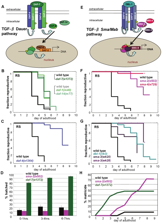
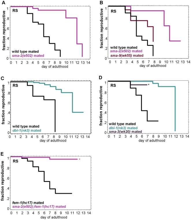
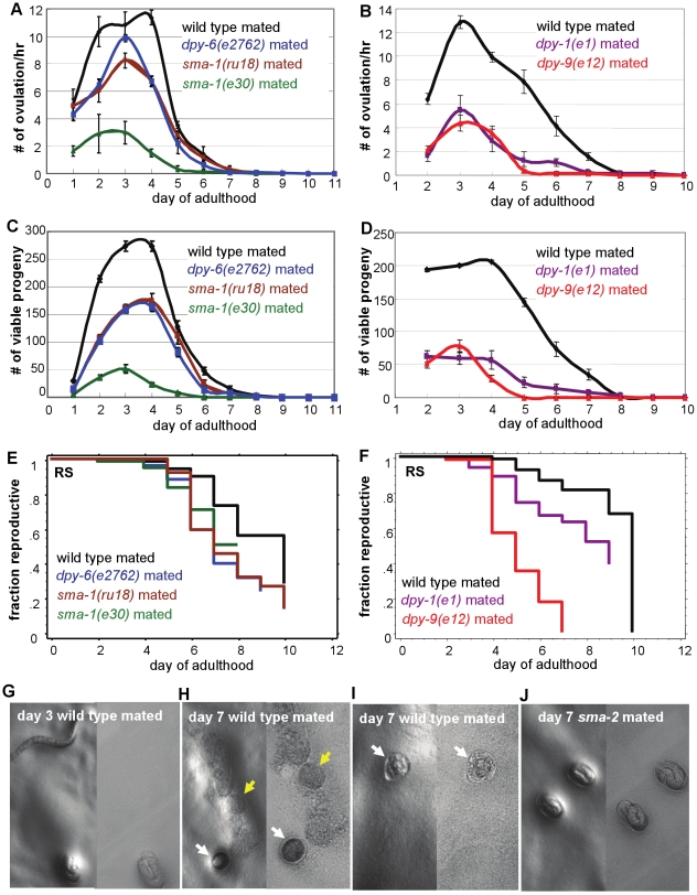
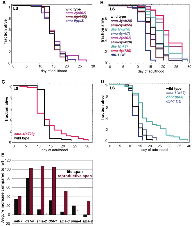
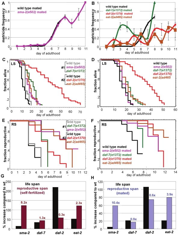
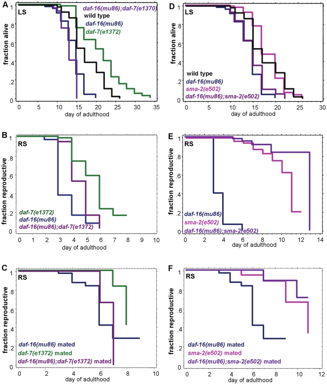
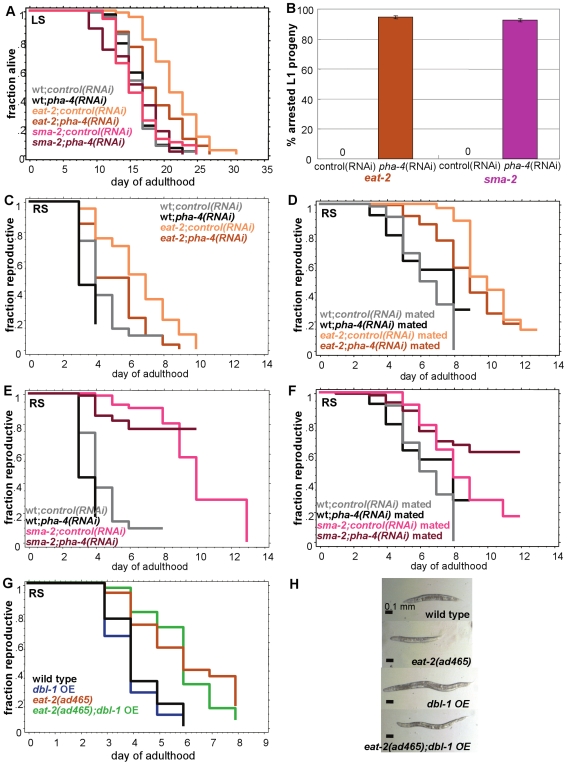
Female reproductive cessation is one of the earliest age-related declines humans experience, occurring in mid-adulthood. Similarly, Caenorhabditis elegans' reproductive span is short relative to its total life span, with reproduction ceasing about a third into its 15-20 day adulthood. All of the known mutations and treatments that extend C. elegans' reproductive period also regulate longevity, suggesting that reproductive span is normally linked to life span. C. elegans has two canonical TGF-beta signaling pathways. We recently found that the TGF-beta Dauer pathway regulates longevity through the Insulin/IGF-1 Signaling (IIS) pathway; here we show that this pathway has a moderate effect on reproductive span. By contrast, TGF-beta Sma/Mab signaling mutants exhibit a substantially extended reproductive period, more than doubling reproductive span in some cases. Sma/Mab mutations extend reproductive span disproportionately to life span and act independently of known regulators of somatic aging, such as Insulin/IGF-1 Signaling and Dietary Restriction. This is the first discovery of a pathway that regulates reproductive span independently of longevity and the first identification of the TGF-beta Sma/Mab pathway as a regulator of reproductive aging. Our results suggest that longevity and reproductive span regulation can be uncoupled, although they appear to normally be linked through regulatory pathways.


Conflict of interest statement

The authors have declared that no competing interests exist.

Figures

Figure 1
Figure 1. TGF-ß Sma/Mab pathway regulates reproductive span.
(A, E) Schematic representation of the TGF-ß Dauer and TGF-ß Sma/Mab pathways in C. elegans. (B) Mutants of the TGF-ß Dauer pathway moderately extend reproductive span (two experiments are shown). (C) A mutant of the shared receptor between the two pathways, daf-4, doubles the reproductive span of wild type. (D) daf-7, but not sma-2, animals lay more-developed progeny than wild type: percentage of embryos hatched 0–1, 3–4, and 6–7 hours after laying from day 3 wild-type, daf-7 and sma-2 adults. (F, G) Mutants of the TGF-ß Sma/Mab pathway greatly extend reproductive span. (H) Cumulative percentage of matricide animals (caused by internal progeny hatching, same below). daf-7's matricide rate is high starting in early adulthood, while sma-2's increases with age, finally resulting in more matricide animals. All reproductive spans in this figure are measured in self-fertilized hermaphrodites. Asterisk indicates high matricide frequency. Additional statistics presented in Table S1 and Table S2.
Figure 2
Figure 2. TGF-ß Sma/Mab mutant reproductive span extensions are independent of sperm contribution.
(A–D) TGF-ß Sma/Mab mutants mated with young wild-type males have significantly longer reproductive spans than mated wild-type animals. (E) After mating with wild-type males, sma-2;fem-1 (spermless) animals also have greater reproductive spans than fem-1 worms. Asterisk indicates a high matricide frequency. Additional statistics presented in Table S1.
Figure 3
Figure 3. Reproductive span is independent of body size, ovulation rate, and progeny number.
Mated non-TGF-ß small mutants all have slower ovulation rates (A, B), and produce fewer early and total progeny (C, D) than wild type. (E, F) Mated reproductive spans of the non-TGF-ß small mutants are not longer than wild type. Additional statistics presented in Table S5. (G–J) Example images of viable embryos (G, J), unhatchable embryos (H, I, white arrows), and unfertilized oocytes (H, yellow arrows) laid by wild type (G–I) or sma-2 (J) mated animals. Images on the left were taken with Leica PLANAPO 1.6× objective, on the right with PLANAPO 5×.
Figure 4
Figure 4. TGF-ß Sma/Mab mutants have larger effects on reproductive span than on life span.
(A–D) Life span assay trials. dbl-1 and sma-6 mutants increase life span moderately (B, D), sma-2 and sma-4 mutants have inconsistent effects on life span (A–C), and sma-3 and sma-9 appear to have no effect on longevity (A, B). dbl-1 OE (over-expression) slightly shortens life span (B, D). (Additional statistics presented in Table S6.) (E) Average percent change from wild-type life span and reproductive span of TGF-ß Dauer and Sma/Mab mutants. Sma/Mab mutants all have larger effects on reproductive span than on life span. Asterisk indicates that the value is not available due to high matricide rate.
Figure 5
Figure 5. TGF-ß Sma/Mab mutants extend reproductive span most disproportionately to longevity effects.
(A, B) Matricide frequencies with age of wild-type, sma-2, daf-7, daf-2, and eat-2 animals (±SEP). Wild-type and sma-2 have similar rates of increasing matricide frequency with age, while daf-2 and eat-2 rates are lower, and daf-7's Egl defect causes very early matricide. (C, D) The life span of sma-2 is shorter than daf-7, daf-2, and eat-2. (E, F) The reproductive span of sma-2 is either longer or comparable to these mutants when self-fertilized (E) or mated (F). (G, H) Percent change from wild-type life span and self-fertilized reproductive span (G) or mated reproductive span (H) of sma-2, daf-7, daf-2, and eat-2. Numbers above reproductive span bars indicate the fold of reproductive span increase over life span increase. sma-2 has the largest effect on reproductive span, compared with its effect on life span. Note that (G) is calculated from (C, E), and (H) is calculated from (D, F). Additional statistics presented in Table S7 and Table S8.
Figure 6
Figure 6. TGF-ß Sma/Mab signaling regulates reproductive span independently of DAF-16/FOXO activity.
(A–C) daf-7's life span (A), self-fertilized reproductive span (B), and mated reproductive span (C) are significantly suppressed by loss of daf-16 activity. (D–F) sma-2's life span (D) is significantly suppressed by daf-16 mutation, but its self-fertilized reproductive span (E) and mated reproductive span (F) are not suppressed by loss of daf-16 activity. (Note that Figure 6A and 6D and Figure 5D are from one experiment; Figure 6C and 6F and Figure 5F are from one experiment.) Additional statistics in Table S9. Matricide data presented in Figure S7B.
Figure 7
Figure 7. TGF-ß Sma/Mab pathway regulates reproductive span independently of dietary restriction.
(A) pha-4 RNAi suppresses eat-2(ad465)'s life span, but has no effect on sma-2(e502)'s life span. (B) sma-2(e502) animals produce similar percentage of arrested L1 progeny as eat-2(ad465) animals when treated with pha-4 RNAi (±SEP, p = 0.17, n = 177 for sma-2, n = 182 for eat-2). When treated with control RNAi, no arrested L1s are produced. (C, D) eat-2(ad465)'s self-fertilized reproductive span (C) and mated reproductive span (D) are both significantly suppressed by pha-4 RNAi treatment. (E, F) Neither sma-2(e502)'s self-fertilized reproductive span (E) nor mated reproductive span (F) are suppressed by loss of pha-4 activity. (Note that (C, E) are from one experiment, while (D, F) are from another.) Additional statistics presented in Table S10. (G) dbl-1 over-expression does not suppress eat-2(ad465)'s reproductive span extension. Additional statistics are in Table S11. (H) Body size of wild type, eat-2(ad465), dbl-1 OE, and eat-2(ad465);dbl-1 OE. eat-2;dbl-1 OE animals are larger than wild type. sma-2;pha-4(RNAi) animals have a severe egg-laying defect, possibly masking even longer reproductive spans (Figure S8).

Similar articles

Cited by

References

    1. Armstrong DT. Effects of maternal age on oocyte developmental competence. Theriogenology. 2001;55:1303–1322. - PubMed
    1. Das S, Blake D, Farquhar C, Seif MMW. Assisted hatching on assisted conception (IVF and ICSI). Cochrane Database of Systematic Reviews. 2009;2:CD001894. - PubMed
    1. Pauli SA, Berga SL, Shang W, Session DR. Current status of the approach to assisted reproduction. Pediatric Clinics of North America. 2009;56:467–488. - PubMed
    1. Palermo GD, Neri QV, Takeuchi T, Rosenwaks Z. ICSI: where we have been and where we are going. Semin Reprod Med. 2009;27:191–201. - PubMed
    1. De Mouzon J. IVF monitoring worldwide (ICMART). Human Reprod. 2006;21:i76.

Publication types

Substances