. 2009 Dec;5(12):e1000708.
doi: 10.1371/journal.ppat.1000708. Epub 2009 Dec 24.

Bacterial effector binding to ribosomal protein s3 subverts NF-kappaB function

Affiliations

Bacterial effector binding to ribosomal protein s3 subverts NF-kappaB function

Xiaofei Gao et al. PLoS Pathog. 2009 Dec.

Abstract

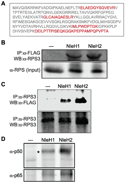
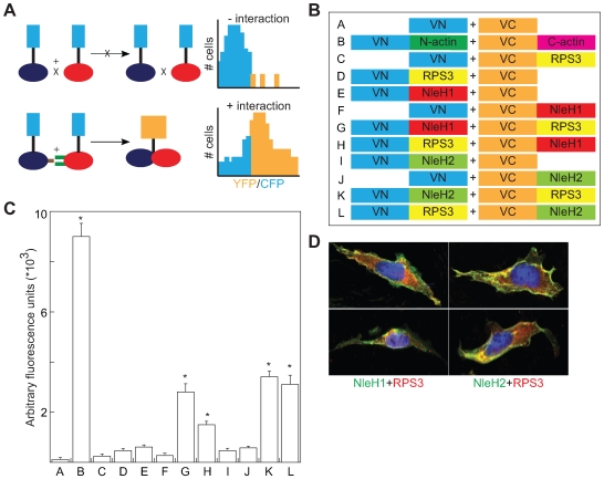
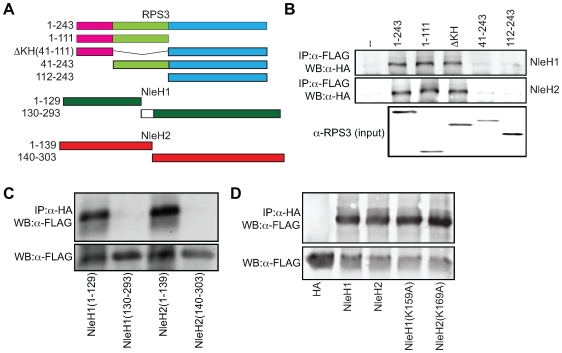
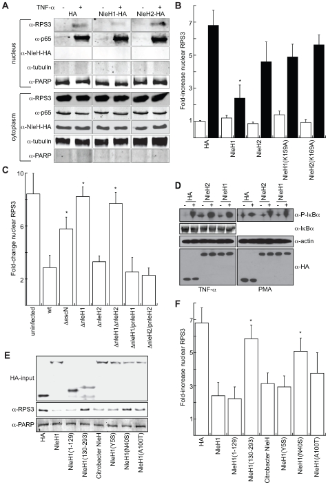
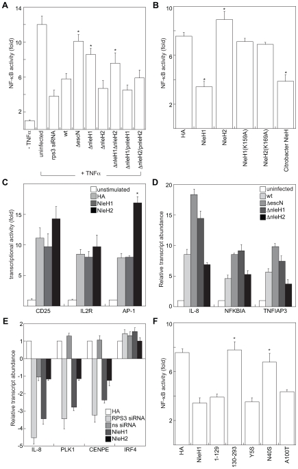
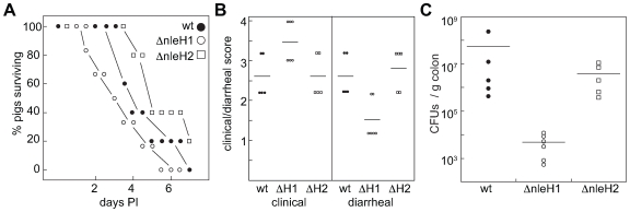
Enteric bacterial pathogens cause food borne disease, which constitutes an enormous economic and health burden. Enterohemorrhagic Escherichia coli (EHEC) causes a severe bloody diarrhea following transmission to humans through various means, including contaminated beef and vegetable products, water, or through contact with animals. EHEC also causes a potentially fatal kidney disease (hemolytic uremic syndrome) for which there is no effective treatment or prophylaxis. EHEC and other enteric pathogens (e.g., enteropathogenic E. coli (EPEC), Salmonella, Shigella, Yersinia) utilize a type III secretion system (T3SS) to inject virulence proteins (effectors) into host cells. While it is known that T3SS effectors subvert host cell function to promote diarrheal disease and bacterial transmission, in many cases, the mechanisms by which these effectors bind to host proteins and disrupt the normal function of intestinal epithelial cells have not been completely characterized. In this study, we present evidence that the E. coli O157:H7 nleH1 and nleH2 genes encode T3SS effectors that bind to the human ribosomal protein S3 (RPS3), a subunit of nuclear factor kappa-light-chain-enhancer of activated B cells (NF-kappaB) transcriptional complexes. NleH1 and NleH2 co-localized with RPS3 in the cytoplasm, but not in cell nuclei. The N-terminal region of both NleH1 and NleH2 was required for binding to the N-terminus of RPS3. NleH1 and NleH2 are autophosphorylated Ser/Thr protein kinases, but their binding to RPS3 is independent of kinase activity. NleH1, but not NleH2, reduced the nuclear abundance of RPS3 without altering the p50 or p65 NF-kappaB subunits or affecting the phosphorylation state or abundance of the inhibitory NF-kappaB chaperone IkappaBalpha NleH1 repressed the transcription of a RPS3/NF-kappaB-dependent reporter plasmid, but did not inhibit the transcription of RPS3-independent reporters. In contrast, NleH2 stimulated RPS3-dependent transcription, as well as an AP-1-dependent reporter. We identified a region of NleH1 (N40-K45) that is at least partially responsible for the inhibitory activity of NleH1 toward RPS3. Deleting nleH1 from E. coli O157:H7 produced a hypervirulent phenotype in a gnotobiotic piglet model of Shiga toxin-producing E. coli infection. We suggest that NleH may disrupt host innate immune responses by binding to a cofactor of host transcriptional complexes.


Conflict of interest statement

The authors have declared that no competing interests exist.

Figures

Figure 1
Figure 1. Translocation and localization of NleH1 and NleH2 in host cells.
A. Immunoblot analysis of cytoplasmic and membrane HeLa cell fractions following infection with EHEC strains expressing NleH1- or NleH2-FLAG fusions. Blots were probed with α-FLAG, α-tubulin, and α-calnexin antibodies. B. Immunofluorescence microscopy analysis of NleH localization. HeLa cells were infected with EHEC ΔescN (left), EHEC/pnleH1-FLAG (middle), or EHEC/pnleH2-FLAG (right) and stained with DAPI (blue) and an α-FLAG monoclonal antibody (green).
Figure 2
Figure 2. NleH1 and NleH2 bind to the host ribosomal protein S3 (RPS3).
A. RPS3 amino acid sequence. Tryptic peptides identified by mass spectrometry are indicated in red. B. Immunoprecipitation of RPS3 from HeLa cells by translocated NleH1- and NleH2-FLAG following infection with EPEC UMD207. Samples were immunoprecipitated with α-FLAG antibody and immunoblotted for RPS3. The lower panel indicates the RPS3 abundance in the cell lysates. C. Immunoprecipitation of NleH1- and NleH2-FLAG from HeLa cells by RPS3 following infection with EPEC UMD207. Samples were immunoprecipitated with α-RPS3 antibody and immunoblotted for FLAG to detect NleH. The lower panel demonstrates the equal enrichment of RPS3 among samples following α-RPS3 immunoprecipitation. D. Enrichment of the p50 and p65 NF-κB subunits by NleH1 and NleH2. Samples were immunoprecipitated with α-FLAG antibody and immunoblotted for p50 (top) and p65 (bottom).
Figure 3
Figure 3. Bimolecular fluorescence complementation analysis of NleH-RPS3 interaction.
A. BiFC schematic. Protein-protein interaction promotes the reconstitution of a functional fluorophore, measured as an increase in the YFP:CFP emission ratio ,. B. Experimental design of NleH- and RPS3-eYFP fusions. VN, N-terminus (AAs 1–173) of Venus fluorescence protein; VC, C-terminus (AAs 155–238) of Venus fluorescence protein. C. Relative fluorescence intensity resulting from the co-transfection of the indicated NleH- and RPS3-eYFP plasmid combinations (n = 3). Asterisks indicate significantly different fluorescence intensity compared with uninfected samples (p<0.05, ANOVA). D. Confocal immunofluorescence microscopy analysis of NleH and RPS3 co-localization. HeLa cells were infected with EPEC strains expressing NleH1- or NleH2-FLAG and stained with DAPI (blue), α-FLAG (green), and α-RPS3 (red) antibodies. Two representative cells are shown for each infection condition.
Figure 4
Figure 4. N-termini of NleH1 and NleH2 bind to the N-terminus of RPS3.
A. Design of RPS3 and NleH protein truncations. The amino acids present in each truncation are indicated to the left of the figure. The death (pink), KH (green), and DNA-repair (blue) domains of RPS3 are indicated. The white box depicts the 10 amino acids lacking in NleH1 but present in NleH2. B. Co-immunoprecipitation of NleH1- (top) and NleH2-HA (middle) with RPS3-FLAG truncations. Samples were immunoprecipitated with α-FLAG antibody to capture RPS3 and immunoblotted for HA to detect NleH. The bottom panel depicts the expression levels of the RPS3-FLAG truncations. C. Co-immunoprecipitation of RPS3-FLAG with NleH1- and NleH2-HA truncations. Samples were immunoprecipitated with α-HA antibody to capture NleH and immunoblotted for FLAG to detect RPS3. The lower panel depicts the expression levels of the NleH truncations. D. Immunoprecipitation of RPS3-FLAG with NleH1(K159A)-HA and NleH2(K169A)-HA. Samples were immunoprecipitated with α-HA antibody to capture NleH and immunoblotted for FLAG to detect RPS3. The lower panel depicts the expression levels of RPS3 in cell lysates.
Figure 5
Figure 5. NleH1 reduces the nuclear abundance of RPS3.
A. Immunoblot analysis of cytoplasmic and nuclear fractions of 293T cells transfected with NleH1, NleH2, or an HA-epitope control, in the presence or absence of TNF-α (100 ng/ml) stimulation for 1 h. Blots were probed with α-RPS3, α-p65, α-HA, α-tubulin, and α-PARP monoclonal antibodies. B. Quantification (n≥4) of the fold-increase in nuclear RPS3 as assessed by densitometry analysis of immunoblots in the absence (open bars) or presence (black bars) of TNF-α stimulation. RPS3 signal intensity was normalized to tubulin (cytoplasmic) and PARP (nuclear). Asterisks indicate significantly different compared with HA transfection (p<0.05, ANOVA). C. Quantification of the fold-increase in nuclear RPS3 following a 3 h infection of HeLa cells with E. coli O157:H7 EDL933 strains possessing or lacking nleH1 and/or nleH2, as well as with strains complemented with the indicated NleH plasmids (n = 3). Asterisks indicate significantly different compared with wild-type infection (p<0.05, ANOVA). D. Immunoblot analysis of IκBα phosphorylation induced by TNF-α (left) or PMA (right), and total IκBα, in the presence or absence of NleH. E. Immunoblot analysis of the impact of C. rodentium NleH and EHEC NleH1 truncations and site-directed mutants on RPS3 nuclear abundance. Blots were probed with α-HA (top), α-RPS (middle), and α-PARP (bottom) monoclonal antibodies. The HA-input panel depicts the expression levels of the indicated constructs. The α-RPS panel depicts the nuclear abundance of RPS3 after stimulation with TNF-α in 293T cells transfected with the indicated constructs. The α-PARP signal was used for normalization of immunoblot signal intensities. F. Quantification (n = 3) of the fold-increase in nuclear RPS3 as assessed by immunoblotting in the presence of the indicated NleH expression plasmids. Asterisks indicate significantly different compared with NleH1 transfection (p<0.05, ANOVA).
Figure 6
Figure 6. NleH effectors alter host NF-κB activity.
A. Relative NF-κB activity (compared with uninfected cells) as a function of TNF-α stimulation, siRNA transfection, and/or infection with EHEC strains possessing or lacking nleH1 and/or nleH2 (n≥4). HeLa cells were co-transfected with a firefly luciferase construct driven by a consensus κB site and a renilla luciferase plasmid, cultured for 36 h, and then infected with EHEC strains for 3 h in the presence or absence of TNF-α stimulation or silencing with rps3 siRNA. Asterisks indicate significantly different compared with wild-type infection (p<0.05, ANOVA). B. Relative NF-κB activity in 293T cells transfected with the indicated NleH plasmids (n = 4). After 36 h, cells were stimulated with TNF-α (100 ng/ml, 1 h). Asterisks indicate significantly different compared with HA transfection (p<0.05, ANOVA). C. Impact of NleH1 and NleH2 on CD25 (left), IL-2R (middle), and AP-1 (right)-dependent luciferase reporter activity. 293T cells were transfected with the indicated reporter plasmids and treated with either TNF-α (CD-25 and IL-2R) or PMA (AP-1) 36 h post-transfection (n = 3). Asterisks indicate significantly different compared with HA transfection (p<0.05, ANOVA). D. Relative transcript abundance, relative to uninfected cells assessed by RT-PCR analysis of 293T cells infected for 4 h with the indicated bacterial strains. IL-8, NFKBIA, and TNFIAP3 data were normalized to GAPDH expression. E. Relative transcript abundance in 293T cells after 48 h transfection with HA, NleH1-HA, NleH2-HA, and RPS3-specific or sequence-scrambled (ns) siRNA constructs. IL-8, PLK1, CENPE, and IRF4 data were normalized to GAPDH expression. F. Impact of C. rodentium NleH and EHEC NleH1 truncations and site-directed mutants on RPS3/NF-κB-dependent transcriptional activity. Experiments were performed as described in panel B, using the plasmids indicated on the x-axis (n = 3). Asterisks indicate significantly different compared with NleH1 transfection (p<0.05, ANOVA).
Figure 7
Figure 7. E. coli O157:H7 ΔnleH1 is hypervirulent in gnotobiotic piglets.
A. Survival analysis of gnotobiotic piglets as a function of time post-inoculation with EHEC strains possessing or lacking nleH1 or nleH2. B. Quantification of piglet clinical outcome (left) and extent of diarrheal disease following EHEC challenge (right). C. Quantification of EHEC colonization of piglet colonic tissue (CFUs/g colon).

References

    1. Clarke SC. Diarrhoeagenic Escherichia coli–an emerging problem? Diagn Microbiol Infect Dis. 2001;41:93–98. - PubMed
    1. Garmendia J, Frankel G, Crepin VF. Enteropathogenic and enterohemorrhagic Escherichia coli infections: translocation, translocation, translocation. Infect Immun. 2005;73:2573–2585. - PMC - PubMed
    1. Donnenberg MS, Kaper JB, Finlay BB. Interactions between enteropathogenic Escherichia coli and host epithelial cells. Trends Microbiol. 1997;5:109–114. - PubMed
    1. Hueck CJ. Type III protein secretion systems in bacterial pathogens of animals and plants. Microbiol Mol Biol Rev. 1998;62:379–433. - PMC - PubMed
    1. McDaniel TK, Jarvis KG, Donnenberg MS, Kaper JB. A genetic locus of enterocyte effacement conserved among diverse enterobacterial pathogens. Proc Natl Acad Sci U S A. 1995;92:1664–1668. - PMC - PubMed

Publication types

MeSH terms