

Platelet activating factor stimulates arachidonic acid release in differentiated keratinocytes via arachidonyl non-selective phospholipase A2
- PMID: 20041255
- PMCID: PMC2829133
- DOI: 10.1007/s00403-009-1017-8
Platelet activating factor stimulates arachidonic acid release in differentiated keratinocytes via arachidonyl non-selective phospholipase A2
Abstract
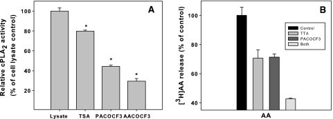
Platelet activating factor (PAF, 1-O-alkyl-2-acetyl-sn-glycero-3-phosphocholine) is known to be present in excess in psoriatic skin, but its exact role is uncertain. In the present study we demonstrate for the first time the role of group VI PLA(2) in PAF-induced arachidonic acid release in highly differentiated human keratinocytes. The group IValpha PLA(2) also participates in the release, while secretory PLA(2)s play a minor role. Two anti-inflammatory synthetic fatty acids, tetradecylthioacetic acid and tetradecylselenoacetic acid, are shown to interfere with signalling events upstream of group IValpha PLA(2) activation. In summary, our major novel finding is the involvement of the arachidonyl non-selective group VI PLA(2) in PAF-induced inflammatory responses.
Figures


Similar articles
-
Distinct roles of two intracellular phospholipase A2s in fatty acid release in the cell death pathway. Proteolytic fragment of type IVA cytosolic phospholipase A2alpha inhibits stimulus-induced arachidonate release, whereas that of type VI Ca2+-independent phospholipase A2 augments spontaneous fatty acid release.J Biol Chem. 2000 Jun 16;275(24):18248-58. doi: 10.1074/jbc.M000271200. J Biol Chem. 2000. PMID: 10747887
-
Hydrolysis of 1-alkyl-2-arachidonoyl-sn-glycero-3-phosphocholine, a common precursor of platelet-activating factor and eicosanoids, by human platelet phospholipase A2.Biochim Biophys Acta. 1988 Apr 15;959(3):269-79. doi: 10.1016/0005-2760(88)90200-7. Biochim Biophys Acta. 1988. PMID: 3355850
-
Expression of phospholipases A2 in primary human lung macrophages: role of cytosolic phospholipase A2-alpha in arachidonic acid release and platelet activating factor synthesis.Biochim Biophys Acta. 2009 Feb;1791(2):92-102. doi: 10.1016/j.bbalip.2008.12.002. Epub 2008 Dec 16. Biochim Biophys Acta. 2009. PMID: 19130898 Free PMC article.
-
Involvement of cytosolic phospholipase A(2), calcium independent phospholipase A(2) and plasmalogen selective phospholipase A(2) in neurodegenerative and neuropsychiatric conditions.Curr Med Chem. 2010;17(25):2746-63. doi: 10.2174/092986710791859289. Curr Med Chem. 2010. PMID: 20586719 Review.
-
Phospholipase A2 biochemistry.Cardiovasc Drugs Ther. 2009 Feb;23(1):49-59. doi: 10.1007/s10557-008-6132-9. Epub 2008 Oct 18. Cardiovasc Drugs Ther. 2009. PMID: 18931897 Free PMC article. Review.
Cited by
-
WFDC12-overexpressing contributes to the development of atopic dermatitis via accelerating ALOX12/15 metabolism and PAF accumulation.Cell Death Dis. 2023 Mar 8;14(3):185. doi: 10.1038/s41419-023-05686-3. Cell Death Dis. 2023. PMID: 36882395 Free PMC article.
-
Platelet-activating factor induces proliferation in differentiated keratinocytes.Mol Cell Biochem. 2013 Dec;384(1-2):83-94. doi: 10.1007/s11010-013-1784-6. Epub 2013 Aug 24. Mol Cell Biochem. 2013. PMID: 23975504
-
Acidification in the epidermis and the role of secretory phospholipases.Dermatoendocrinol. 2011 Apr;3(2):84-90. doi: 10.4161/derm.3.2.15140. Epub 2011 Apr 1. Dermatoendocrinol. 2011. PMID: 21695017 Free PMC article.
References
-
- Anthonsen MW, Andersen S, Solhaug A, Johansen B. Atypical lambda/iota PKC conveys 5-lipoxygenase/leukotriene B4-mediated cross-talk between phospholipase A2 s regulating NF-kappa B activation in response to tumor necrosis factor-alpha and interleukin-1beta. J Biol Chem. 2001;276(38):35344–35351. doi: 10.1074/jbc.M105264200. - DOI - PubMed
Publication types
MeSH terms
Substances
LinkOut - more resources
Full Text Sources
