

Neonatal maternal separation disrupts regulation of sleep and breathing in adult male rats
- PMID: 20041597
- PMCID: PMC2786045
- DOI: 10.1093/sleep/32.12.1611
Neonatal maternal separation disrupts regulation of sleep and breathing in adult male rats
Abstract
Study objectives: Neonatal maternal separation (NMS) disrupts development of cardiorespiratory regulation. Adult male rats previously subjected to NMS are hypertensive and show a hypoxic ventilatory response greater than that of controls. These results have been obtained in awake or anesthetised animals, and the consequences of NMS on respiratory control during normal sleep are unknown. This study tested the following.
Hypotheses: NMS augments respiratory variability across sleep-wake states, and NMS-related enhancement of the hypoxic ventilatory response occurs during sleep.
Methods: Two groups of adult rats were used: controls (no treatment) and rats subjected to NMS. Ventilatory activity, coefficient of variation, and hypoxic ventilatory response were compared between groups and across sleep-wake states.
Subjects: Male Sprague Dawley rats-NMS: n=11; controls: n=10. Pups subjected to NMS were isolated from their mother for 3 hours per day from postnatal days 3 to 12. Controls were undisturbed.
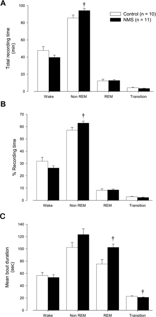
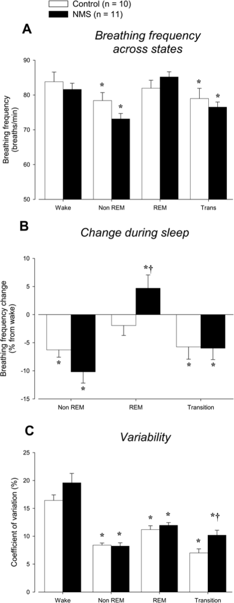
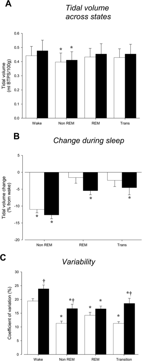
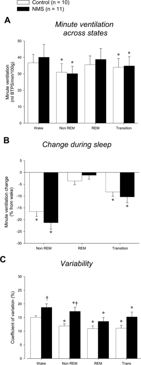
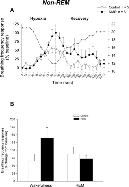
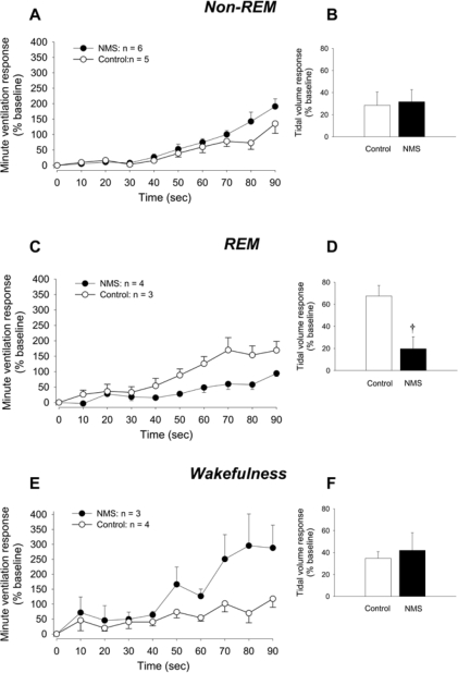
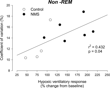
Measurements and results: At adulthood, sleep-wake states were monitored by telemetry, and ventilatory activity was measured using whole-body plethysmography. Sleep and breathing were measured for 2.5 hours (in the morning) while the rats were breathing room air. Data were analysed in 20-second epochs. Rats were then exposed to a brief (90-sec) hypoxic episode (nadir = 12% O2) to measure the hypoxic ventilatory response. The coefficient of variability for tidal volume and breathing frequency decreased during sleep but remained more elevated in NMS rats than in controls. During non-rapid eye movement sleep, the breathing-frequency response to hypoxia of NMS rats was significantly greater than that of controls.
Conclusion: Neonatal maternal separation results in persistent disruption of respiratory control during sleep.
Figures
Similar articles
-
Testosterone potentiates the hypoxic ventilatory response of adult male rats subjected to neonatal stress.Exp Physiol. 2014 May 1;99(5):824-34. doi: 10.1113/expphysiol.2013.077073. Epub 2014 Mar 7. Exp Physiol. 2014. PMID: 24610832
-
Neonatal stress affects the aging trajectory of female rats on the endocrine, temperature, and ventilatory responses to hypoxia.Am J Physiol Regul Integr Comp Physiol. 2015 Apr 1;308(7):R659-67. doi: 10.1152/ajpregu.00418.2014. Epub 2015 Feb 4. Am J Physiol Regul Integr Comp Physiol. 2015. PMID: 25652536
-
Neonatal maternal separation and sex-specific plasticity of the hypoxic ventilatory response in awake rat.J Physiol. 2004 Jan 15;554(Pt 2):543-57. doi: 10.1113/jphysiol.2003.052894. Epub 2003 Nov 21. J Physiol. 2004. PMID: 14634199 Free PMC article.
-
Neonatal maternal separation and neuroendocrine programming of the respiratory control system in rats.Biol Psychol. 2010 Apr;84(1):26-38. doi: 10.1016/j.biopsycho.2009.09.001. Epub 2009 Sep 6. Biol Psychol. 2010. PMID: 19737597 Review.
-
Intermittent hypoxia, respiratory plasticity and sleep apnea in humans: present knowledge and future investigations.Respir Physiol Neurobiol. 2013 Sep 15;188(3):289-300. doi: 10.1016/j.resp.2013.04.010. Epub 2013 Apr 12. Respir Physiol Neurobiol. 2013. PMID: 23587570 Free PMC article. Review.
Cited by
-
Exposure to early adversity: Points of cross-species translation that can lead to improved understanding of depression.Dev Psychopathol. 2015 May;27(2):477-91. doi: 10.1017/S0954579415000103. Dev Psychopathol. 2015. PMID: 25997766 Free PMC article. Review.
-
Estrogens, age, and, neonatal stress: panic disorders and novel views on the contribution of non-medullary structures to respiratory control and CO2 responses.Front Physiol. 2023 May 17;14:1183933. doi: 10.3389/fphys.2023.1183933. eCollection 2023. Front Physiol. 2023. PMID: 37265841 Free PMC article. Review.
-
Maternal opioids age-dependently impair neonatal respiratory control networks.Front Physiol. 2023 Mar 16;14:1109754. doi: 10.3389/fphys.2023.1109754. eCollection 2023. Front Physiol. 2023. PMID: 37008014 Free PMC article.
-
Effect of Estrous Cycle on Behavior of Females in Rodent Tests of Anxiety.Front Psychiatry. 2021 Aug 31;12:711065. doi: 10.3389/fpsyt.2021.711065. eCollection 2021. Front Psychiatry. 2021. PMID: 34531768 Free PMC article. Review.
-
Sex, hormones, and stress: how they impact development and function of the carotid bodies and related reflexes.Respir Physiol Neurobiol. 2013 Jan 1;185(1):75-86. doi: 10.1016/j.resp.2012.07.001. Epub 2012 Jul 8. Respir Physiol Neurobiol. 2013. PMID: 22781657 Free PMC article. Review.
References
-
- Rietveld S, van Beest I, Everaerd W. Stress-induced breathlessness in asthma. Psychol Med. 1999;29:1359–66. - PubMed
-
- Gunnar MR. Integrating neuroscience and psychological approaches in the study of early experiences. Ann N Y Acad Sci. 2003;1008:238–47. - PubMed
-
- Seckl JR, Meaney MJ. Early life events and later development of ischaemic heart disease. The Lancet. 1993;342:1236. - PubMed
-
- Nicolson NA. Childhood parental loss and cortisol levels in adult men. Psychoneuroendocrinology. 2004;29:1012–8. - PubMed
Publication types
MeSH terms
Grants and funding
LinkOut - more resources
Full Text Sources
Medical
