. 2010 Jan 21;114(2):952-8.
doi: 10.1021/jp906790f.

Ion selectivity of alpha-hemolysin with a beta-cyclodextrin adapter. I. Single ion potential of mean force and diffusion coefficient

Affiliations

Ion selectivity of alpha-hemolysin with a beta-cyclodextrin adapter. I. Single ion potential of mean force and diffusion coefficient

Yun Luo et al. J Phys Chem B. .

Abstract

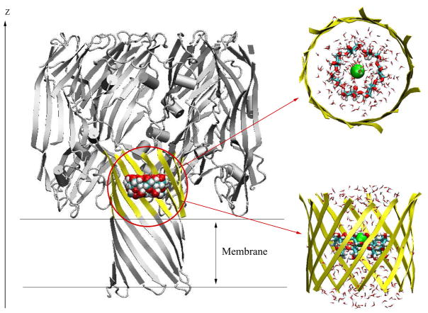
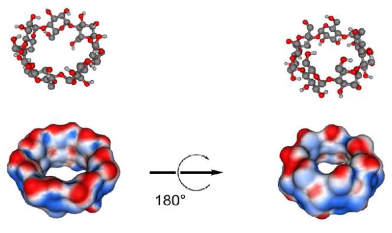
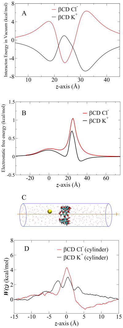
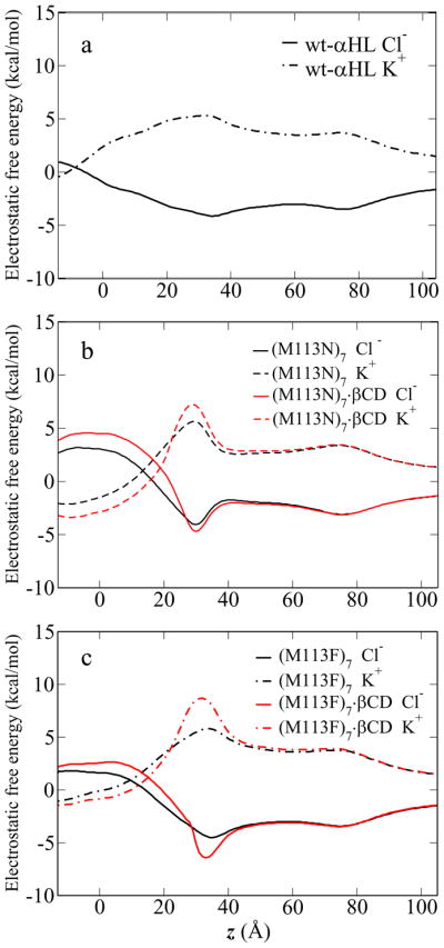
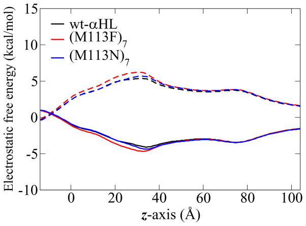
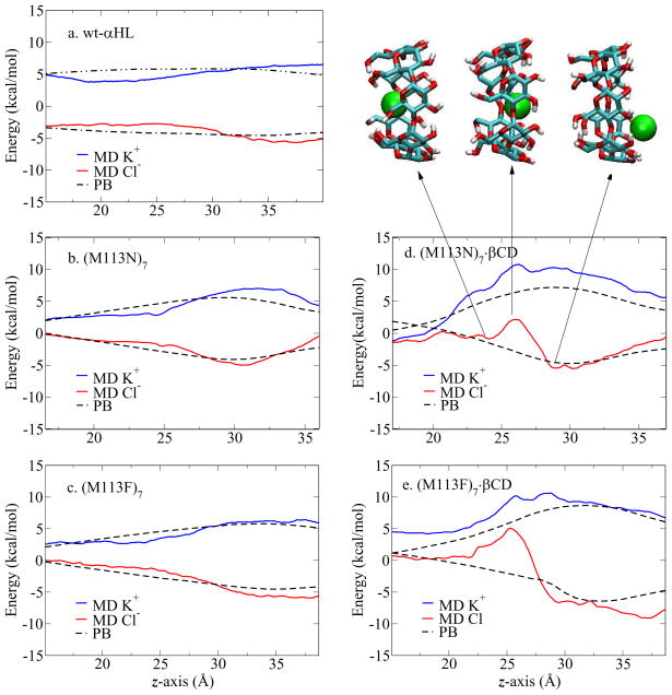
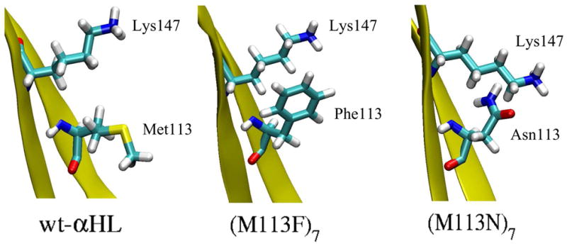
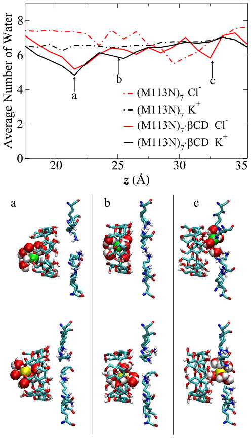
The alpha-hemolysin (alphaHL) is a self-assembling exotoxin that binds to the membrane of a susceptible host cell and causes its death. Experimental studies show that electrically neutral beta-cyclodextrin (betaCD) can insert into the alphaHL channel and significantly increase its anion selectivity. To understand how betaCD can affect ion selectivity, molecular dynamics simulations and potential of mean force (PMF) calculations are carried out for different alphaHL channels with and without the betaCD adapter. A multiscale approach based on the generalized solvent boundary potential is used to reduce the size of the simulated system. The PMF profiles reveal that betaCD has no anion selectivity by itself but can increase the Cl(-) selectivity of the alphaHL channel when lodged into the pore lumen. Analysis shows that betaCD causes a partial desolvation of ions and affects the orientation of nearby charged residues. The ion selectivity appears to result from increased electrostatic interaction between the ion and the channel due to a reduction in dielectric shielding by the solvent. These observations suggest a reasonable explanation of the ion selectivity and provide important information for further ion channel modification.


Figures

Figure 1
Figure 1
Molecular graphics view of the GSBP simulation system for (M113N)7 channel with βCD adapter inside. The full protein is shown in cartoon style (grey), while the inner GSBP simulation region is highlighted in yellow. Channel axis is along the z-axis. Implicit membrane is centered at z = 0 Å with thickness of 25 Å. The reduced system for GSBP is zoomed in and shown in top view and side view. The residues in the inner region, yellow ribbons; βCD, VDW model in whole channel and licorice model in zoom-in figure; Cl, green ball; TIP3 water, stick in atom type colors.
Figure 2
Figure 2
Pore radius profiles along the channel axis calculated using CHARMM program. wt-αHL in black, (M113F)7 in red and (M113N)7 in blue.
Figure 3
Figure 3
β-cyclodextrin is shown as ball-and-stick models and as Connolly surfaces onto which electrostatic potential is mapped (blue = positive, red = negative). Partial atomic charges and force field parameters were calculated using the General Amber Force Field/AM1/BCC method.
Figure 4
Figure 4
(A) Interaction of K+ and Cl ions with βCD in vacuum. βCD is located at z = 25 Å. The ion is moving from 5 Å to 45 Å through βCD along the z-axis. (B) The electrostatic free energy profiles of ion passage through βCD, obtained from PB calculations. βCD is located at z = 25 Å. (C) The cartoon of single ion passage through βCD in the cylinder of explicit water solvent. βCD, licorice in atom type colors; K+ in yellow ball; TIP3 water in red stick. (D) 1D-PMF of K+and Cl passage through βCD in the cylinder of explicit water solvent.
Figure 5
Figure 5
The electrostatic free energy profiles of ion passage through five channels, obtained from PB calculations. The solid lines are Cl profiles and the dashed lines are K+ profile.
Figure 6
Figure 6
The electrostatic free energy profiles obtained from PB calculations. The (M113N)7 and (M113F)7 mutants are re-constructed from the wt-αHL X-ray structure, with the configuration of the other side chains unchanged. The solid lines are Cl profiles and the dashed lines are K+ profile.
Figure 7
Figure 7
1D-PMF profiles of ion passage through five channels. In PMF profiles, the red line is Cl profile, the blue line is K+ profile and the dashed line is the electrostatic free energy profile. Three snapshots show the Cl position corresponding to the z coordinates in the PMF profile. βCD is in licorice with atom type colors; Cl in green ball; protein and TIP3 water molecules are not shown here.
Figure 8
Figure 8
The snapshots of partial segment of channels showing the orientation of Lys147 and the residue at position 113 in three crystal structures. The (M113F)7 has Lys147 in the same orientation as the wt-αHL. While in the (M113N)7 Lys147 is pointing slightly downwards.
Figure 9
Figure 9
Average number of water molecules in the first solvation shell of ion inside (M113N)7 and (M113N)7·βCD channels. Radius cutoff 3.7 Å is used for Cl (red lines), 3.5 Å for K+ (black lines). Snapshots show the typical ion and water configurations inside (M113N)7·βCD channel at ion position of (a) z ~ 21 Å, (b) z ~ 25 Å, (c) z ~ 33 Å. The three positions are also pointed out in the figure. Cl in green VDW sphere; K+ in yellow VDW sphere; TIP3P water in VDW sphere with atom type colors; both βCD and Lys147 residues are shown in licorice style with atom type colors. Only water molecules within the first hydration shell are shown here, other residues and water molecules are omitted for clarity.

Similar articles

Cited by

References

    1. Laestadius A, Richter-Dahlfors A, Aperia A. Kidney Int. 2002;62:2035–2042. - PubMed
    1. Gentschev I, Dietrich G, Goebel W. Trends in Microbiology. 2002;10:39–45. - PubMed
    1. Rowe SM, Miller S, Sorscher EJ. New England Journal of Medicine. 2005;352:1992–2001. - PubMed
    1. Gouaux E. J Struct Biol. 1998;121:110–122. - PubMed
    1. Aksimentiev A, Schulten K. Biophys J. 2005;88:3745–3761. - PMC - PubMed

Publication types