Review
. 2010;16(8):942-55.
doi: 10.2174/138161210790883426.

Echinoderms: potential model systems for studies on muscle regeneration

Affiliations
Review

Echinoderms: potential model systems for studies on muscle regeneration

José E García-Arrarás et al. Curr Pharm Des. 2010.

Abstract

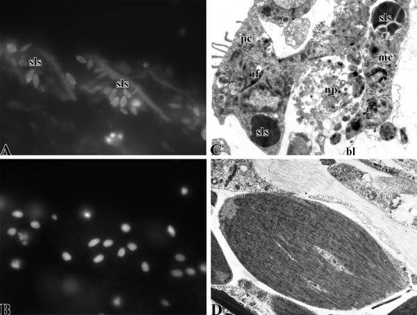
Organisms of the phylum Echinodermata show some of the most impressive regenerative feats within the animal kingdom. Following injury or self-induced autotomy, species in this phylum can regenerate most tissues and organs, being the regeneration of the muscular systems one of the best studied. Even though echinoderms are closely related to chordates, they are little known in the biomedical field, and therefore their uses to study pharmacological effects on muscle formation and/or regeneration have been extremely limited. In order to rectify this lack of knowledge, we describe here the echinoderm muscular systems, particularly the somatic and visceral muscle components. In addition, we provide details of the processes that are known to take place during muscle regeneration, namely dedifferentiation, myogenesis and new muscle formation. Finally, we provide the available information on molecular and pharmacological studies that involve echinoderm muscle regeneration. We expect that by making this information accessible, researchers consider the use of echinoderms as model systems for pharmacological studies in muscle development and regeneration.


Figures

Fig. (1)
Fig. (1)
Phylogenetic tree showing the relationship of echinoderms to other animal groups.
Fig. (2)
Fig. (2)
Schemes of normal organization of muscle systems in echinoderms. A. Visceral musculature (coelomic epithelium) is composed of ciliated peritoneal cells with bundles of intermediate filaments (green) and groups of myoepithelial cells (green-reddish). The latter contain myofilaments (red spots and lines). Moreover the epithelium contains nerve cells and their processes (blue). Peritoneal and myoepithelial cells adhere to basal lamina (deep brown) by hemidesmosomes. The coelomic epithelium is situated on connective tissue layer (brown). B. Muscle bundle of somatic musculature. The muscle bundle is composed of several myocytes (pink) and surrounded by basal lamina (deep brown). The cytoplasm of the cell contains myofilaments (red spots and lines). Besides myocytes, muscle bundles contain putative neurons and their processes (blue).
Fig. (3)
Fig. (3)
Dedifferentiation of cell of visceral musculature. A. Spindle-like structures (sls) and fibers labeled with rhodamine-labeled phalloidin in regenerating intestinal mesentery. B. Cell nuclei labeled with DAPI. Note the lack of correlation between SLS and cell nuclei. C. Electron micrograph of regenerating cloacal coelomic epithelium during regeneration of respiratory trees. Note fragmented bundles of intermediate filaments (if) in peritoneal cells (pc). The cells loose connections to the basal lamina (bl). SLS (sls) can be observed in the cytoplasm of both peritoneocytes and myoepithelial cells (mc). There are bundles of nerve processes (np). D. Electron micrograph of SLS.
Fig. (4)
Fig. (4)
Disappearance of muscle cells from the mesentery in animals regenerating their intestine. A. Few muscle cells are labeled with a muscle-specific antibody in the mesentery of a specimen undergoing intestinal regeneration one week following evisceration. B. In contrast, the mesentery of a normal non-regenerating animal shows well-organized muscle system labeled with the same antibody.
Fig. (5)
Fig. (5)
Dedifferentiation of myocytes of somatic musculature. A. Large numbers of SLSs are found in the muscle stump 6-days following transection of longitudinal muscle band of H. glaberrima. B. Higher magnification shows the details of the SLSs among disorganized muscle fibers. C. Electron micrograph of apoptotic cells (ac) within in the connective tissue (ct) of the muscle stump 3-days after transection of longitudinal muscle band of E. fraudatrix. D. Electron micrograph of the myocyte with fragmented myofilament-containing cytoplasm (mf). Note activated nucleus (n) of the myocyte.
Fig. (6)
Fig. (6)
Regeneration of visceral musculature. A. Electron micrograph of the differentiating myocyte situated under peritoneal cells (pc) in regenerating respiratory tree. Note bundle of nerve processes (n) and growing process of myocyte with bundle of myofilaments (mf). B-E. The process of muscle formation can be followed using a muscle-specific antibody in the regenerating intestine of H. glaberrima. Cells in the coelomic epithelium express the muscle epitope during the first week of regeneration (B, F). By the second week (C, G) myocytes (me) are found beneath the peritoneocytes (ce). By the third week (D, H) large numbers of myocytes are found although their orientation is not evident. Four weeks into regeneration (E, I) the two muscle layers, longitudinal (lm) and circular (cm) are evident.
Fig. (7)
Fig. (7)
Regeneration of somatic musculature. A. Electron micrograph of group of coelomic epithelial cells sinking into the connective tissue (ct) of the longitudinal muscle band of E. fraudatrix. Note cell nuclei (n) and long processes containing bundles of myofilaments (mf) adjacent to the coelomic cavity (cc). B. Electron micrograph of differentiating myocytes (dm). Their cytoplasm contains bundles of myofilaments (mf), and well-developed rough endoplasmic reticulum (rer) and Golgi apparatus (ga).
Fig. (8)
Fig. (8)
Schemes of dedifferentiation and regeneration of muscle system in echinoderms. A. Transformation of the coelomic epithelium during regeneration. a. Organization of the coelomic epithelium. This epithelium is formed of ciliated peritoneal cells with bundles of intermediate filaments (green) and groups of myoepithelial cells (green-reddish) with myofilaments (red spots and lines). b. Dedifferentiation of the coelomic epithelial cells begins just after damaging. Bundles of intermediate filaments are destroyed, peritoneal cells loose connection with basal lamina (brown). Spindle-like structures are formed in myoepithelial cells. Some of them are exocytosed into the coelomic cavity or endocytosed by peritoneal cells. c. Partial dedifferentiation of the coelomic epithelial cells. (As occurs during regeneration of respiratory trees in holothurians.) Dedifferentiated peritoneal cells have lost connections with basal lamina but remain attached to other by cell junctions. These cells can undergo mitosis. Dedifferentiated myoepithelial cells form a mesenchymal layer under the peritoneal cells. These cells do not contain myofilaments and do not divide. d. Full dedifferentiation of coelomic epithelium. (As occurs during gut regeneration in holothurians.) Peritoneal and myoepithelial cells intermingle to form a single epithelium on basal lamina. Cells of the epithelium divide mitotically. e. Beginning of redifferentiation. Some cells give rise to peritoneal cells and other – myoepithelial cells. Cytoplasm of the latter contains small bundles of myofilaments. f. Redifferentiation of the coelomic epithelium. Peritoneal cells form basal processes and attach to basal lamina by hemidesmosomes. Their cytoplasm contains small bundles of intermediate filaments. Myoepithelial cells are situated under peritoneal cells and develop long myofilament- containing processes. B. Regeneration of longitudinal muscle band in holothurians. a. Undamaged muscle band composed of bundles of muscles cells (red) and covered by coelomic epithelium containing only peritoneal cells (green). Muscle band is situated on connective tissue layer (brown) of body wall. b. Muscle band following transection. c. Beginning of regeneration. Coelomic epithelium migrates to wound region and covers it. Damaged myocytes are destroyed. d. Formation of new muscle bundles (green-reddish ovals). Groups of peritoneal cells sink into connective tissue and form muscle bundle. Their cytoplasm contains myofilaments. Concurrently, destruction of several muscle bundles in the wound region continues. Myocytes dedifferentiate. They shed myofilament-containing cytoplasm and the cell transforms into a myoblast. e. Advanced stage of regeneration. Coelomic epithelium continues formatting new muscle bundles. Concurrently, groups of myoblasts begin to form new muscle bundle (reddish oval). f. Regenerated muscle band contains old bundles (red) and new bundles develop from coelomic epithelium (green-reddish ovals) so dedifferentiated myocytes (reddish ovals). g. Higher magnification of region 1 shows destruction of muscle bundles. h. Higher magnification of region 2 shows formation of new muscle bundle (nm) and old muscle bundles (om) with dedifferentiating myocytes. i. Higher magnification of region 3 shows formation of new muscle bundle (nm), old muscle bundles (om) with dedifferentiating myocytes, and muscle bundle regenerated from dedifferentiated myocytes (rm).
Fig. (9)
Fig. (9)
Muscle regeneration in crinoids. A. Semi thin section of normal muscle (M) in crinoid arm. B. Dedifferentiating muscle in crinoid arm subjected to 2.5 μg/ml of the antiandrogenic drug DDE for 72 hrs. Muscle dedifferentiation is increased in the presence of various drugs (mainly endocrine disruptors). The drugs used were: (a) Triphenyltin (b) fenarimol (c) methyl-testosterone and (d) p, p’-DDE. Increases in muscle dedifferentiation cause a decrease in the length of the regenerating arm length as observed by the effect of various doses of the endocrine disruptor drugs (modified from Sugni et al. 2007, 2008).

References

    1. Sánchez Alvarado A, Tsonis PA. Bridging the regeneration gap: genetic insight from diverse animal models. Nature Rev Genet. 2006;7:873–83. - PubMed
    1. Brockes JP, Kumar A. Comparative aspects of animal regeneration. Annu Rev Cell Dev Biol. 2008;24:525–49. - PubMed
    1. Adoutte A, Balavoine G, Lartillot N, Lespinet O, Prud'homme B, de Rosa R. The new animal phylogeny: Reliability and implications. Proc Natl Acad Sci USA. 2000;97:4453–56. - PMC - PubMed
    1. Blair JE, SE, Hedges S. Molecular phylogeny and divergence times of deuterostome animals. Mol Biol Evol. 2005;22:22–75-84. - PubMed
    1. Swalla BJ, Smith AB. Deciphering deuterostome phylogeny: molecular, morphologoical and palaentological perspectives. Phil Trans R Soc B. 2008;363:1557–68. - PMC - PubMed

Publication types

Substances