Disorders related with ageing in the gerbil female prostate (Skene's paraurethral glands)
- PMID: 20041966
- PMCID: PMC2965899
- DOI: 10.1111/j.1365-2613.2009.00685.x
Disorders related with ageing in the gerbil female prostate (Skene's paraurethral glands)
Abstract
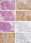
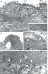
The female organs, which are regulated by steroid hormones, are targets of studies especially those related to senescence. However, although the female prostate is an organ influenced by hormones and susceptible to lesions, there is still little information about its histopathology. Thus, given the morphophysiological similarity between the prostate in women and female gerbils, the present study aimed to identify the spontaneous histopathological changes in this rodent to provide contributions to the understanding of lesions that also affect the human female prostate. The structural, ultrastructural, immunohistochemical, morphometric-stereological and serological aspects, as well as the quantification of the incidence, multiplicity and percentage of acini affected by different lesions were analyzed. Benign prostate lesions including hyperplasia, prostatitis, microcalculi and calculi; preneoplastic lesions like dysplasias; premalignant lesions, such as high grade prostatic intraepithelial neoplasia as well as malignant ones, specifically adenocarcinoma, were identified in the adult gland, but they were intensified during senescence, which is possibly due to the imbalance among steroid hormone levels. Although clinical attention focuses on other urogenital organs, the real condition of the histopathological injuries in the human female prostate should be considered. A serious preventive work regarding the female prostate could be applied in the gynaecological context in order to monitor the gland and avoid possible disturbances to women's health and consequently provide better quality of life.
Figures


Similar articles
-
Aging effects on the mongolian gerbil female prostate (Skene's paraurethral glands): structural, ultrastructural, quantitative, and hormonal evaluations.Anat Rec (Hoboken). 2008 Apr;291(4):463-74. doi: 10.1002/ar.20637. Anat Rec (Hoboken). 2008. PMID: 18231985
-
Combined oral contraceptives promote androgen receptor and oestrogen receptor alpha upregulation in the female prostate (Skene's paraurethral glands) of adult gerbils (Meriones unguiculatus).Reprod Fertil Dev. 2018 Oct;30(10):1286-1297. doi: 10.1071/RD17294. Reprod Fertil Dev. 2018. PMID: 29622059
-
Microscopic evaluation of proliferative disorders in the gerbil female prostate: evidence of aging and the influence of multiple pregnancies.Micron. 2011 Oct;42(7):712-7. doi: 10.1016/j.micron.2011.03.011. Epub 2011 Apr 8. Micron. 2011. PMID: 21531142
-
The female prostate and prostate-specific antigen. Immunohistochemical localization, implications of this prostate marker in women and reasons for using the term "prostate" in the human female.Histol Histopathol. 2000 Jan;15(1):131-42. doi: 10.14670/HH-15.131. Histol Histopathol. 2000. PMID: 10668204 Review.
-
Skene's gland adenocarcinoma with intestinal differentiation: A case report and literature review.Pathol Int. 2017 Nov;67(11):575-579. doi: 10.1111/pin.12571. Epub 2017 Sep 5. Pathol Int. 2017. PMID: 28872768 Review.
Cited by
-
Exposure to ethinylestradiol during prenatal development and postnatal supplementation with testosterone causes morphophysiological alterations in the prostate of male and female adult gerbils.Int J Exp Pathol. 2011 Apr;92(2):121-30. doi: 10.1111/j.1365-2613.2010.00756.x. Epub 2011 Feb 12. Int J Exp Pathol. 2011. PMID: 21314741 Free PMC article.
-
Female Hyperplastic PSA-Positive Prostate Tissue in the Bladder Neck.Urol Int. 2024;108(2):163-167. doi: 10.1159/000535605. Epub 2023 Dec 6. Urol Int. 2024. PMID: 38056438 Free PMC article.
-
Prenatal exposure to ethinylestradiol alters the morphologic patterns and increases the predisposition for prostatic lesions in male and female gerbils during ageing.Int J Exp Pathol. 2016 Feb;97(1):5-17. doi: 10.1111/iep.12153. Epub 2016 Feb 8. Int J Exp Pathol. 2016. PMID: 26852889 Free PMC article.
-
Telocytes play a key role in prostate tissue organisation during the gland morphogenesis.J Cell Mol Med. 2017 Dec;21(12):3309-3321. doi: 10.1111/jcmm.13234. Epub 2017 Aug 24. J Cell Mol Med. 2017. PMID: 28840644 Free PMC article.
References
-
- Ali SZ, Smilari TF, Gal D, Lovecchio JL, Teichberg S. Primary adenoid cystic carcinoma of Skene′s glands. Gynecol. Oncol. 1995;57:257–261. - PubMed
-
- Bonkhoff H, Remberger K. Morphogenetic concepts of normal and abnormal growth in the human prostate. Virchows Arch. 1998;433:195–202. - PubMed
-
- Brawer MK. Prostatic intraepithelial neoplasia: a premalignant lesion. Symposium: the pathology of prostate cancer – prt 1: classic aspects of prostate cancer pathology. Hum. Pathol. 1992;23:242–248. - PubMed
-
- Carson C, Rittmaster R. The role of dihydrotestosterone in benign prostatic hyperplasia. Urology. 2003;61:2–7. - PubMed
-
- Chahal HS, Drake WM. The endocrine system and ageing. J. Pathol. 2007;21:173–180. - PubMed
Publication types
MeSH terms
Substances
LinkOut - more resources
Full Text Sources
Medical

