

The principled control of false positives in neuroimaging
- PMID: 20042432
- PMCID: PMC2799957
- DOI: 10.1093/scan/nsp053
The principled control of false positives in neuroimaging
Abstract
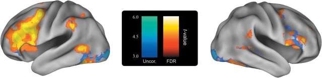
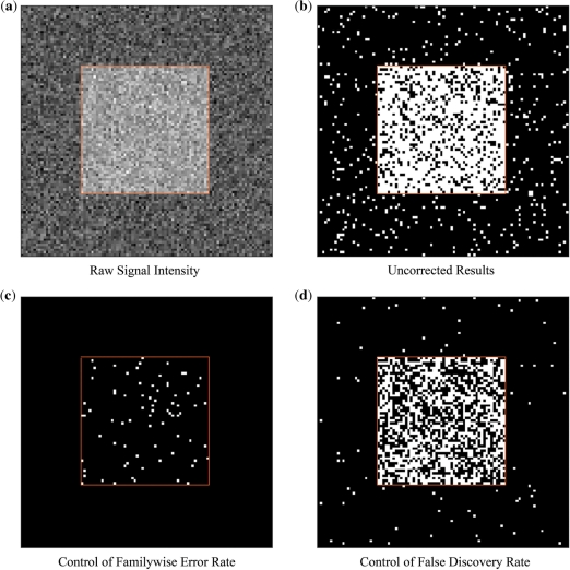
An incredible amount of data is generated in the course of a functional neuroimaging experiment. The quantity of data gives us improved temporal and spatial resolution with which to evaluate our results. It also creates a staggering multiple testing problem. A number of methods have been created that address the multiple testing problem in neuroimaging in a principled fashion. These methods place limits on either the familywise error rate (FWER) or the false discovery rate (FDR) of the results. These principled approaches are well established in the literature and are known to properly limit the amount of false positives across the whole brain. However, a minority of papers are still published every month using methods that are improperly corrected for the number of tests conducted. These latter methods place limits on the voxelwise probability of a false positive and yield no information on the global rate of false positives in the results. In this commentary, we argue in favor of a principled approach to the multiple testing problem--one that places appropriate limits on the rate of false positives across the whole brain gives readers the information they need to properly evaluate the results.
Figures
References
-
- Arnott SR, Cant JS, Dutton GN, Goodale MA. Crinkling and crumpling: an auditory fMRI study of material properties. Neuroimage. 2008;43(2):368–78. - PubMed
-
- Baker CI, Hutchison TL, Kanwisher N. Does the fusiform face area contain subregions highly selective for nonfaces? Nature Neuroscience. 2007;10(1):3–4. - PubMed
-
- Benjamini Y, Hochberg Y. “Controlling the false discovery rate: A practical and powerful approach to multiple testing”. Journal of the Royal Statistic Society Series B. 1995;57:289–300.
-
- Bennett CM, Baird AA, Miller MB, Wolford GL. 15th Annual Meeting of the Organization for Human Brain Mapping. San Francisco, CA: 2009. Neural correlates of interspecies perspective taking in the post-mortem atlantic salmon: an argument for proper multiple comparisons correction.
Publication types
MeSH terms
LinkOut - more resources
Full Text Sources
Medical
