. 2010 Feb;16(2):394-404.
doi: 10.1261/rna.1905910. Epub 2009 Dec 30.

Anti-Argonaute RIP-Chip shows that miRNA transfections alter global patterns of mRNA recruitment to microribonucleoprotein complexes

Affiliations

Anti-Argonaute RIP-Chip shows that miRNA transfections alter global patterns of mRNA recruitment to microribonucleoprotein complexes

Wang-Xia Wang et al. RNA. 2010 Feb.

Abstract

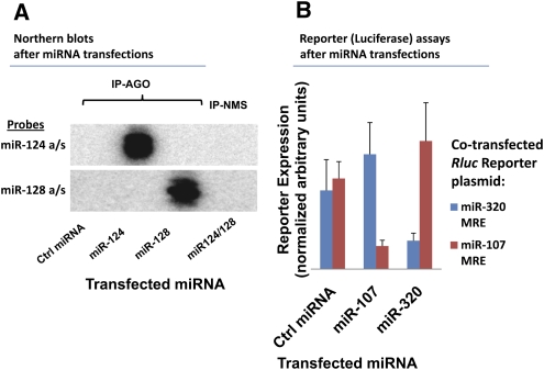
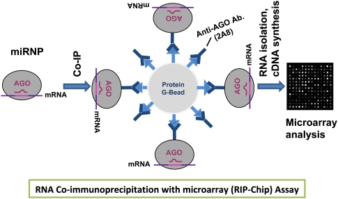
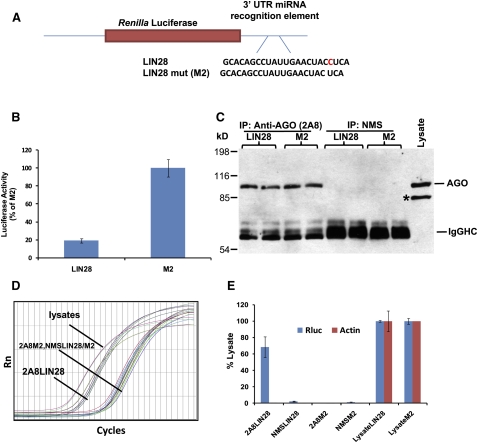
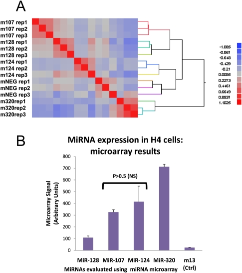
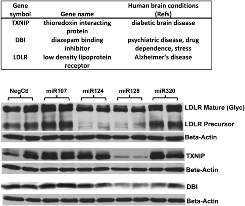
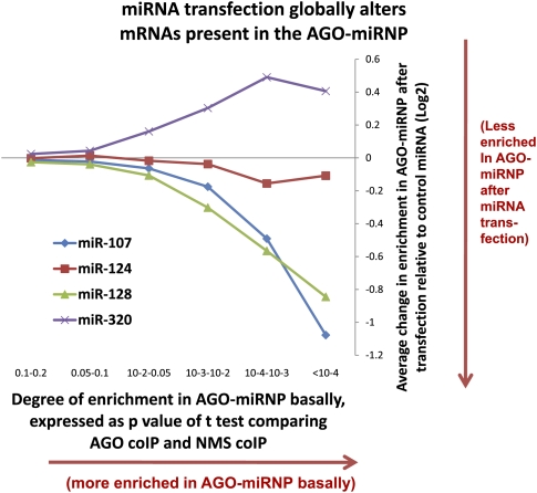
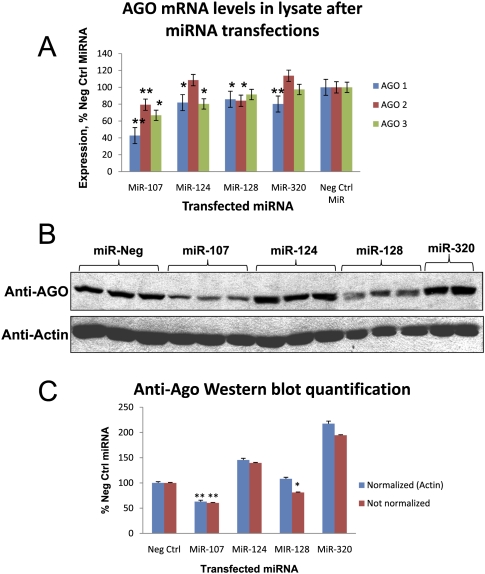
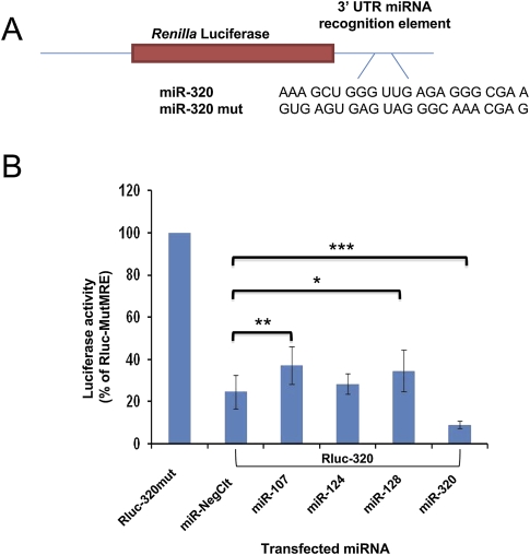
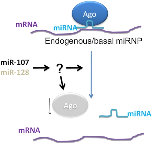
MicroRNAs (miRNAs) play key roles in gene expression regulation by guiding Argonaute (AGO)-containing microribonucleoprotein (miRNP) effector complexes to target polynucleotides. There are still uncertainties about how miRNAs interact with mRNAs. Here we employed a biochemical approach to isolate AGO-containing miRNPs from human H4 tumor cells by co-immunoprecipitation (co-IP) with a previously described anti-AGO antibody. Co-immunoprecipitated (co-IPed) RNAs were subjected to downstream Affymetrix Human Gene 1.0 ST microarray analysis. During rigorous validation, the "RIP-Chip" assay identified target mRNAs specifically associated with AGO complexes. RIP-Chip was performed after transfecting brain-enriched miRNAs (miR-107, miR-124, miR-128, and miR-320) and nonphysiologic control miRNA to identify miRNA targets. As expected, the miRNA transfections altered the mRNA content of the miRNPs. Specific mRNA species recruited to miRNPs after miRNA transfections were moderately in agreement with computational target predictions. In addition to recruiting mRNA targets into miRNPs, miR-107 and to a lesser extent miR-128, but not miR-124 or miR-320, caused apparent exclusion of some mRNAs that are normally associated with miRNPs. MiR-107 and miR-128 transfections also result in decreased AGO mRNA and protein levels. However, AGO mRNAs were not recruited to miRNPs after either miR-107 or miR-128 transfection, confirming that miRNAs may alter gene expression without stable association between particular mRNAs and miRNPs. In summary, RIP-Chip assays constitute an optimized, validated, direct, and high-throughput biochemical assay that provides data about specific miRNA:mRNA interactions, as well as global patterns of regulation by miRNAs.


Figures

FIGURE 1.
FIGURE 1.
MicroRNA (miRNA) transfections are effective and lead to specific targeting by miRNAs. All transfections (25 nM) were performed in H4 glioneuronal cells and cells were harvested 48 h after transfection. (A) Northern blot analysis of miRNPs isolated after miRNA transfections with miR-124 and miR-128. The Northern blot analysis showed that miRNAs were specifically recruited to miRNPs after miRNA transfections. (B) Renilla luciferase (Rluc) reporter assays showed that miRNAs are functionally incorporated after miRNA transfections. MiR-107 and miR-320 specifically target reporter constructs bearing 3′-untranslated region recognition sequences for those miRNAs.
FIGURE 2.
FIGURE 2.
Schematic illustration of biochemical identification of miRNA targets using RIP-Chip. miRNPs are co-IPed with anti-AGO (2A8) antibodies (Nelson et al. 2007) bound to protein G-agarose beads. RNAs that associated with AGO protein are processed for Affymetrix Gene 1.0 ST microarrays and this high-throughput assay can resolve miRNA targets.
FIGURE 3.
FIGURE 3.
RIP-Chip was validated in H4 cells using positive miRNA target control and closely matched negative controls with transfected Renilla luciferase (Rluc) reporter plasmids. (A) Let-7 miRNA recognition element (MRE) of LIN28 or its closely related mutated sequence (designated “M2”) in the 3′-untranslated region (UTR) of the Rluc gene as previously described (Kiriakidou et al. 2004). (B) Dual luciferase activity assay shows that LIN28 reporter activity was decreased as expected by endogenous Let-7 miRNA. (C) Western blots analysis of co-IPed products using anti-AGO antibody. As expected, AGO proteins were co-IPed with anti-AGO (2A8), but not with nonimmunized mouse serum (NMS). Co-IPs were performed on cells transfected with reporter plasmid Rluc-LIN28 or Rluc-M2. An asterisk (*) shows a band that is Radixin, a protein recognized on Western blots, but not via co-IP by anti-AGO 2A8. (D) Although AGO protein was co-IPed regardless of the transfected plasmids, RT-qPCR detected Rluc mRNA only in the anti-AGO co-IP from the cell lysates transfected with Rluc-LIN28. (E) Quantification of qPCR shows that Rluc mRNA was highly enriched only in co-IPed RNAs from cells transfected with Rluc-LIN28, demonstrating a highly specific association of Rluc mRNA with AGO proteins via miRNA binding sites.
FIGURE 4.
FIGURE 4.
(A) Clustering analysis of RIP-Chip microarray expression data indicating that upstream miRNA transfections cause consistent changes in the mRNAs recruited into the miRNP. Anti-AGO co-IPs of H4 cells were processed for Affymetrix Gene 1.0 ST microarrays. This heat-map representation of the microarray expression profiling after transfection with various miRNAs and RIP-Chip comprises 15 different biological replicates. The abscissa refers to the same 15 biological replicates in the same order. The analysis indicates that the transfections produced consistent results with between-group variability exceeding within-group variability. (B) Endogenous expression levels of selected miRNAs in H4 cells. Levels of selected brain enriched miRNAs were evaluated as described previously (Wang et al. 2008b) by miRNA microarray analysis of H4 cell RNAs.
FIGURE 5.
FIGURE 5.
Western blot analysis after miRNA transfections helped validate RIP-Chip results. H4 cells were first transfected with full-length cDNA plasmids: pCMV6-LDLR, pCMV6-TXNIP, and pCMV6-DBI for 24 h, and were then transfected with 25 nM of miR-107, miR-124, miR-128, miR-320 precursors, or a negative control miRNA for additional 48 h. Cells were harvested, and Western blot analyses were performed using antibodies against each expressing proteins. Anti-β-actin antibody was used to probe each blot to monitor the total protein loadings. The three miR-128 targeted proteins shown were effectively knocked down by miR-128 transfection. LDLR was also strongly reduced by miR-124 transfection. RIP-Chip did not identify LDLR as miR-124 target, however, microarray analysis of total cell RNA showed that LDLR mRNA was significant decreased upon miR-124 transfection.
FIGURE 6.
FIGURE 6.
MiRNA transfections lead to systematic changes in the mRNAs that are incorporated into AGO-miRNPs as determined by RIP-Chip. The abscissa shows six different groups of mRNAs. Enrichment in the AGO-miRNP increases from left to right according to the P-value of t-tests comparing the mRNA amount in anti-AGO co-IP versus anti-NMS co-IP. mRNA groups were P = 0.1–0.2 (N = 3503 mRNAs), P = 0.05–0.1 (N = 1786), P = 0.01–0.05 (N = 1594), P = 0.001–0.01 (N = 531), P = 0.00001–0.0001 (N = 82), and P < 0.00001 (N = 10). Note that for the group with the greatest enrichment in the AGO-miRNPs at baseline (right-most data point), transfections with miR-107 and miR-128, but not miR-124 or miR-320, cause a decrease in these mRNAs in the AGO-miRNP.
FIGURE 7.
FIGURE 7.
MiR-107 and miR-128 transfections lead to reduced AGO expression. (A) Microarray analysis of the lysates prior to co-IP shows that different miRNA transfections differentially alter the level of AGO mRNAs. Note that miR-107 and miR-128 transfections cause AGO2 mRNAs to decrease. “*,” P < 0.05; “**,” P < 0.01; using Student's t-test, n = 3 replicates each. (B) Western blot analysis of AGO protein expression after H4 cells were transfected with 25 nM of various miRNAs. Total cell proteins were subjected to SDS-PAGE followed by Western blot analysis using anti-AGO antibody, or anti-β-actin antibody. (C) In a separate experiment, three biological replicates of miRNA transfections were performed for each miRNA and Western blot analysis using anti-AGO and anti-actin. AGO and β-actin protein levels of each individual band were quantified blindly on the Western blot and then normalized to levels of β-actin. Note that AGO protein was knocked down by miR-107 and miR-128. Although equal amounts of total protein were loaded in each well, β-actin levels seemed to be reduced in cells following miR-128 transfections.
FIGURE 8.
FIGURE 8.
Reporter assays are used to test how endogenous miRNA/miRNP function is affected by exogenous miRNA transfections. H4 cells were first transfected with various miRNA precursors (negative control, miR-107, miR-124, miR-128, and miR-320) for 48 h A. Reporter plasmid constructs pRL-TK-Rluc carrying miR-320 MRE (Rluc-320), or its mutated sequence (Rluc-320mut) were co-transfected with calibrating plasmid pGL3 for an additional 24 h B. Luciferase activities were determined as previously described (Wang et al. 2008a,b). Luciferase activities, normalized to PGL-3 values, from each transfection were compared to that of negative control miRNA which represents endogenous miR-320 activity. Represented are three independent experiments each with three replicates. Student's t-tests were performed on the data set. “*,” P < 0.05; “**,” P < 0.01; “***,” P < 0.001.
FIGURE 9.
FIGURE 9.
Cartoon schematic to show that miR-107 and miR-128 transfections decrease the enrichment of endogenous mRNA targets in AGO-miRNPs. MiR-107 and miR-128 also lead to decreased miRNA/miRNP activity by reporter assays, correlated with decreased AGO mRNA and protein in the cells. However, since AGO mRNAs are not enriched in miRNPs, the mechanism for the down-regulation of AGO is as yet unknown.

Similar articles

Cited by

References

    1. Adams BD, Claffey KP, White BA. Argonaute-2 expression is regulated by epidermal growth factor receptor and mitogen-activated protein kinase signaling and correlates with a transformed phenotype in breast cancer cells. Endocrinology. 2009;150:14–23. - PMC - PubMed
    1. Ambros V. The functions of animal microRNAs. Nature. 2004;431:350–355. - PubMed
    1. Andachi Y. A novel biochemical method to identify target genes of individual microRNAs: Identification of a new Caenorhabditis elegans let-7 target. RNA. 2008;14:2440–2451. - PMC - PubMed
    1. Arnstein P, Taylor DO, Nelson-Rees WA, Huebner RJ, Lennette EH. Propagation of human tumors in antithymocyte serum-treated mice. J Natl Cancer Inst. 1974;52:71–84. - PubMed
    1. Baek D, Villen J, Shin C, Camargo FD, Gygi SP, Bartel DP. The impact of microRNAs on protein output. Nature. 2008;455:64–71. - PMC - PubMed

Publication types

MeSH terms