. 2010 Mar;84(6):2893-901.
doi: 10.1128/JVI.01616-09. Epub 2009 Dec 30.

Epstein-Barr virus-encoded Bcl-2 homologue functions as a survival factor in Wp-restricted Burkitt lymphoma cell line P3HR-1

Affiliations

Epstein-Barr virus-encoded Bcl-2 homologue functions as a survival factor in Wp-restricted Burkitt lymphoma cell line P3HR-1

Ami Watanabe et al. J Virol. 2010 Mar.

Abstract

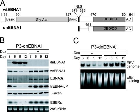
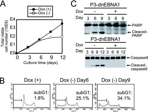
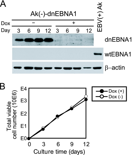
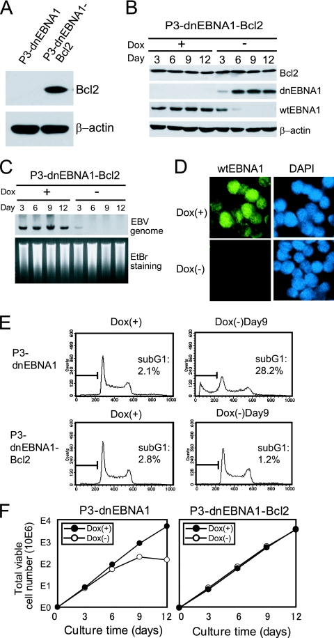
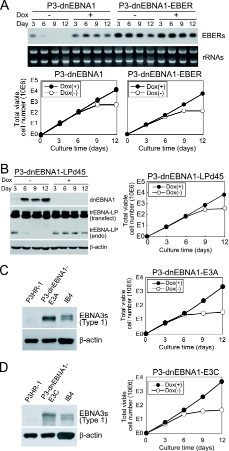
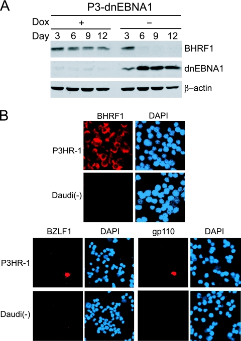
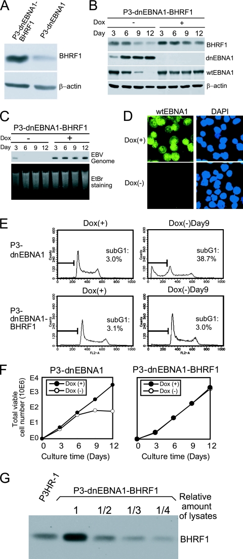
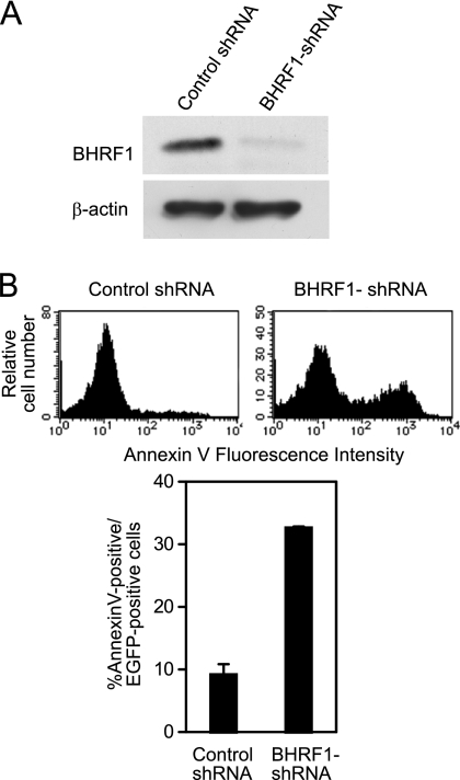
Burkitt lymphoma (BL) is etiologically associated with Epstein-Barr virus (EBV). EBV-positive BL tumors display two latent forms of infection. One is referred to as latency I infection, in which EBV expresses the virus genome maintenance protein EBNA1 as the only viral protein. The other is referred to as Wp-restricted latency and was recently identified in a subset of BL tumors. In these tumors, EBV expresses EBNA1, EBNA3A, EBNA3B, EBNA3C, a truncated form of EBNA-LP, and the viral Bcl-2 homologue BHRF1, all of which are driven by the BamHI W promoter (Wp). To investigate the role of EBV in Wp-restricted BL, we conditionally expressed a dominant-negative EBNA1 (dnEBNA1) mutant which interrupts the virus genome maintenance function of EBNA1 in the P3HR-1 BL cell line. Induction of dnEBNA1 expression caused loss of the EBV genome and resulted in apoptosis of P3HR-1 cells in the absence of exogenous apoptosis inducers, indicating that P3HR-1 cells cannot survive without EBV. Stable transfection of the BHRF1 gene into P3HR-1 cells rescued the cells from the apoptosis induced by dnEBNA1 expression, whereas stable transfection of truncated EBNA-LP, EBNA3A, or EBNA3C did not. Moreover, knockdown of BHRF1 expression in P3HR-1 cells resulted in increased cell death. These results indicate that EBV is essential for the survival of P3HR-1 cells and that BHRF1 functions as a survival factor. Our finding implies a critical contribution of BHRF1 to the pathogenesis of Wp-restricted BLs.


Figures

FIG. 1.
FIG. 1.
Enforced expression of dnEBNA1 in P3HR-1 cells results in loss of the EBV genome and viral gene products. (A) Schematic representations of wt EBNA1 and dnEBNA1. The basic amino acid-rich chromosome association domains (Basic), the Gly-Gly-Ala repeats (Gly-Ala), the nuclear localization sequence (NLS), the DNA-binding and dimerization domain (DBD/DD), and the acidic domain (AC) are shown. The numbers indicate the positions of amino acid residues. (B) P3-dnEBNA1 cells were cultured in the absence (−) or presence (+) of Dox. Every 3 days, the cultures were split. Total cell lysates were prepared at the indicated time points and subjected to Western blot analysis with EBV-immune human sera (reactive to dnEBNA1, wt EBNA1, or EBNA3s), anti-EBNA-LP (reactive to truncated [tr] EBNA-LP), or anti-β-actin antibody (β-actin). Total RNAs were prepared at the indicated time points and subjected to Northern blot analysis to detect EBERs. Ethidium bromide-stained 28S rRNA as a loading control is shown below. (C) P3-dnEBNA1 cells were cultured in the absence (−) or presence (+) of Dox. Every 3 days, the cultures were split. Genomic DNAs were prepared at the indicated time points, digested with EcoRI, and subjected to Southern blot analysis to detect the EBV genome. Ethidium bromide-stained DNA as a loading control is shown below (EtBr staining).
FIG. 2.
FIG. 2.
Enforced expression of dnEBNA1 in P3HR-1 cells results in the suppression of cell growth and the induction of apoptosis. (A) P3-dnEBNA1 cells were cultured in the absence (−) or presence (+) of Dox. Every 3 days, viable cells were counted, and the cells were split as described in Materials and Methods. The total numbers of viable cells derived from the initial cultures were calculated and plotted at each time point. The number “10E6” along the y axis indicates that the cell number plotted should be multiplied by 1,000,000. (B) P3-dnEBNA1 cells were cultured in the absence (−) or presence (+) of Dox for the indicated number of days. The cells were fixed and stained with propidium iodide, and cell cycle analysis was performed with a FACSCalibur. The percentages of apoptotic cells were determined and are shown (sub-G1). (C) P3-dnEBNA1 cells were cultured in the absence (−) or presence (+) of Dox. Total cell lysates were prepared at the indicated time points and subjected to Western blot analysis with anti-PARP (top) or anti-caspase 9 (bottom) antibody.
FIG. 3.
FIG. 3.
Enforced expression of dnEBNA1 does not affect the growth of EBV-negative Akata cells. (A) Ak(−)-dnEBNA1 cells were cultured in the absence (−) or presence (+) of Dox. Total cell lysates were prepared and subjected to Western blot analysis with EBV-immune human serum (reactive to dnEBNA1 and wt EBNA1) and anti-β-actin antibody. EBV-positive Akata cells served as a positive control for wt EBNA1 detection [EBV(+)Ak]. (B) Ak(−)-dnEBNA1 cells were cultured in the absence (−) or presence (+) of Dox. Viable cells were counted every 3 days, and the cells were split. The total numbers of viable cells derived from the initial cultures were calculated and plotted.
FIG. 4.
FIG. 4.
Stable overexpression of Bcl-2 rescues P3HR-1 cells from dnEBNA1-induced growth inhibition and apoptosis. (A) P3-dnEBNA1 and P3-dnEBNA1-Bcl2 cells were subjected to Western blot analysis with anti-Bcl-2 and anti-β-actin antibodies. (B) P3-dnEBNA1-Bcl2 cells cultured in the absence (−) or presence (+) of Dox were subjected to Western blot analysis with EBV-immune human serum (reactive to dnEBNA1 and wt EBNA1), anti-Bcl-2, and anti-β-actin antibodies. (C) P3-dnEBNA1-Bcl2 cells cultured in the absence (−) or presence (+) of Dox were subjected to Southern blot analysis to detect the EBV genome. (D) P3-dnEBNA1-Bcl2 cells were cultured in the absence (−) or presence (+) of Dox for 12 days. The cells were processed for immunofluorescence analyses with EBV-immune human serum (reactive to wt EBNA1, but not to dnEBNA1) as a primary antibody and FITC-conjugated anti-C3C antibody as a secondary antibody. (E) P3-dnEBNA1 cells and P3-dnEBNA1-Bcl2 cells were cultured in the absence (−) or presence (+) of Dox for 9 days. Cell cycle analysis was performed, and the percentages of cells in the sub-G1 fraction were determined. (F) P3-dnEBNA1 and P3-dnEBNA1-Bcl2 cells were cultured in the absence (−) or presence (+) of Dox. Viable cells were counted every 3 days, and the cells were split. The total numbers of viable cells derived from the initial cultures were calculated and plotted.
FIG. 5.
FIG. 5.
Stable transfection with EBERs, truncated EBNA-LP, EBNA3A, or EBNA3C does not protect P3HR-1 cells from dnEBNA1-induced growth inhibition. (A) P3-dnEBNA1 and P3-dnEBNA1-EBER cells cultured in the absence (−) and presence (+) of Dox were subjected to Northern blot analysis to detect EBERs. An ethidium bromide-stained rRNA loading control is also shown. Viable cells were counted, and the total numbers of viable cells derived from the initial cultures were calculated and plotted. (B) P3-dnEBNA1-LPd45 cells were subjected to Western blot analysis with EBV-immune human serum (reactive to dnEBNA1), anti-EBNA-LP, and anti-β-actin antibodies (left). The bands representing transfected truncated (tr) EBNA-LP (transfect) and endogenous trEBNA-LP (endo) are shown. Viable cells were counted, and the total numbers of viable cells derived from the initial cultures were calculated and plotted (right). (C and D) P3HR-1, P3-dnEBNA1-E3A, and P3-dnEBNA1-E3C cells were subjected to Western blot analysis with human serum (reactive to type 1 EBNA3s, but not to type 2 EBNA3s) and anti-β-actin antibody. Note that transfected EBNA3s are type 1 and P3HR-1s endogenous EBNA3s are type 2. An IB4 lysate was used as a positive control for type 1 EBNA3A and type 1 EBNA3C (left). Viable cells were counted, and the total numbers of viable cells derived from the initial cultures were calculated and plotted (right).
FIG. 6.
FIG. 6.
Expression of BHRF1 as a latent protein in P3HR-1 cells. (A) P3-dnEBNA1 cells cultured in the absence (−) or presence (+) of Dox were subjected to Western blot analysis with EBV-immune human serum (reactive to dnEBNA1), anti-BHRF1, and anti-β-actin antibodies. (B) P3HR-1 cells and EBV-negative Daudi cells [Daudi(−)] were processed for immunofluorescence analyses with mouse anti-BHRF1, rabbit anti-BZLF1, or mouse anti-gp110 antibody as a primary antibody and Cy3-conjugated anti-mouse IgG or Cy3-conjugated anti-rabbit IgG as a secondary antibody.
FIG. 7.
FIG. 7.
Stable transfection of the BHRF1 gene protects P3HR-1 cells from dnEBNA1-induced apoptosis and growth inhibition. (A) P3-dnEBNA1 and P3-dnEBNA1-BHRF1 cells cultured in medium with Dox were subjected to Western blot analysis with anti-BHRF1 and anti-β-actin antibodies. (B) P3-dnEBNA1-BHRF1 cells cultured in the absence (−) or presence (+) of Dox were subjected to Western blot analysis with EBV-immune human serum (reactive to dnEBNA1 and wt EBNA1), anti-BHRF1, and anti-β-actin antibodies. (C) P3-dnEBNA1-BHRF1 cells cultured in the absence (−) or presence (+) of Dox were subjected to Southern blot analysis to detect the EBV genome. (D) P3-dnEBNA1-BHRF1 cells were cultured in the absence (−) or presence (+) of Dox for 12 days. The cells were processed for immunofluorescence analyses with EBV-immune human serum (reactive to wt EBNA1, but not to dnEBNA1) as a primary antibody and FITC-conjugated anti-C3C antibody as a secondary antibody. (E) P3-dnEBNA1 and P3-dnEBNA1-BHRF1 cells were cultured in the absence (−) or presence (+) of Dox for 9 days. Cell cycle analysis was performed, and the percentages of cells in the sub-G1 fraction were determined. (F) P3-dnEBNA1 and P3-dnEBNA1-BHRF1 cells were cultured in the absence (−) or presence (+) of Dox. Viable cells were counted every 3 days, and the cells were split. The total numbers of viable cells derived from the initial cultures were calculated and plotted. (G) Cell lysates of P3HR-1 and serially diluted cell lysates of P3-dnEBNA1-BHRF1 were subjected to Western blot analysis with anti-BHRF1 antibody.
FIG. 8.
FIG. 8.
Knockdown of BHRF1 expression in P3HR-1 cells results in increased cell death. (A) P3HR-1 cells were transfected with oriP plasmid expressing BHRF1-shRNA (or control shRNA) and EGFP. Three days after transfection, EGFP-positive cells were sorted and subjected to Western blot analysis with anti-BHRF1 and anti-β-actin antibodies. (B) P3HR-1 cells transfected with BHRF1-shRNA (or control shRNA) and EGFP were stained with annexin V-PE 4 days after transfection. The histograms represent annexin V staining after gating on EGFP-positive cells (top). The percentages of annexin V-positive cells among EGFP-positive cells are shown (bottom). Each bar represents the average of triplicate transfections, with the error bar representing the standard error of the mean.

Similar articles

Cited by

References

    1. Anderton, E., J. Yee, P. Smith, T. Crook, R. E. White, and M. J. Allday. 2008. Two Epstein-Barr virus (EBV) oncoproteins cooperate to repress expression of the proapoptotic tumour-suppressor Bim: clues to the pathogenesis of Burkitt's lymphoma. Oncogene 27:421-433. - PubMed
    1. Askew, D. S., R. A. Ashmun, B. C. Simmons, and J. L. Cleveland. 1991. Constitutive c-myc expression in an IL-3-dependent myeloid cell line suppresses cell cycle arrest and accelerates apoptosis. Oncogene 6:1915-1922. - PubMed
    1. Austin, P. J., E. Flemington, C. N. Yandava, J. L. Strominger, and S. H. Speck. 1988. Complex transcription of the Epstein-Barr virus BamHI fragment H rightward open reading frame 1 (BHRF1) in latently and lytically infected B lymphocytes. Proc. Natl. Acad. Sci. U. S. A. 85:3678-3682. - PMC - PubMed
    1. Bissonnette, R. P., F. Echeverri, A. Mahboubi, and D. R. Green. 1992. Apoptotic cell death induced by c-myc is inhibited by bcl-2. Nature 359:552-554. - PubMed
    1. Bornkamm, G. W., J. Hudewentz, U. K. Freese, and U. Zimber. 1982. Deletion of the nontransforming Epstein-Barr virus strain P3HR-1 causes fusion of the large internal repeat to the DSL region. J. Virol. 43:952-968. - PMC - PubMed

Publication types

MeSH terms

Substances

LinkOut - more resources