The capsid proteins of Aleutian mink disease virus activate caspases and are specifically cleaved during infection
- PMID: 20042496
- PMCID: PMC2826067
- DOI: 10.1128/JVI.01917-09
The capsid proteins of Aleutian mink disease virus activate caspases and are specifically cleaved during infection
Abstract
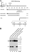
Aleutian mink disease virus (AMDV) is currently the only known member of the genus Amdovirus in the family Parvoviridae. It is the etiological agent of Aleutian disease of mink. We have previously shown that a small protein with a molecular mass of approximately 26 kDa was present during AMDV infection and following transfection of capsid expression constructs (J. Qiu, F. Cheng, L. R. Burger, and D. Pintel, J. Virol. 80:654-662, 2006). In this study, we report that the capsid proteins were specifically cleaved at aspartic acid residue 420 (D420) during virus infection, resulting in the previously observed cleavage product. Mutation of a single amino acid residue at D420 abolished the specific cleavage. Expression of the capsid proteins alone in Crandell feline kidney (CrFK) cells reproduced the cleavage of the capsid proteins in virus infection. More importantly, capsid protein expression alone induced active caspases, of which caspase-10 was the most active. Active caspases, in turn, cleaved capsid proteins in vivo. Our results also showed that active caspase-7 specifically cleaved capsid proteins at D420 in vitro. These results suggest that viral capsid proteins alone induce caspase activation, resulting in cleavage of capsid proteins. We also provide evidence that AMDV mutants resistant to caspase-mediated capsid cleavage increased virus production approximately 3- to 5-fold in CrFK cells compared to that produced from the parent virus AMDV-G at 37 degrees C but not at 31.8 degrees C. Collectively, our results indicate that caspase activity plays multiple roles in AMDV infection and that cleavage of the capsid proteins might have a role in regulating persistent infection of AMDV.
Figures


Similar articles
-
Internal polyadenylation of parvoviral precursor mRNA limits progeny virus production.Virology. 2012 May 10;426(2):167-77. doi: 10.1016/j.virol.2012.01.031. Epub 2012 Feb 21. Virology. 2012. PMID: 22361476 Free PMC article.
-
Molecular characterization of the small nonstructural proteins of parvovirus Aleutian mink disease virus (AMDV) during infection.Virology. 2014 Mar;452-453:23-31. doi: 10.1016/j.virol.2014.01.005. Epub 2014 Jan 28. Virology. 2014. PMID: 24606679 Free PMC article.
-
Identification and characterization of a novel B-cell epitope on Aleutian Mink Disease virus capsid protein VP2 using a monoclonal antibody.Virus Res. 2018 Mar 15;248:74-79. doi: 10.1016/j.virusres.2017.12.008. Epub 2017 Dec 24. Virus Res. 2018. PMID: 29278728
-
AMDV Vaccine: Challenges and Perspectives.Viruses. 2021 Sep 14;13(9):1833. doi: 10.3390/v13091833. Viruses. 2021. PMID: 34578415 Free PMC article. Review.
-
[Eenie, Meenie, Miney, Moe, who is responsible for the antibody-dependent enhancement of Aleutian mink disease parvovirus infection?].Bing Du Xue Bao. 2014 Jul;30(4):450-5. Bing Du Xue Bao. 2014. PMID: 25272602 Review. Chinese.
Cited by
-
Parvovirus infection-induced cell death and cell cycle arrest.Future Virol. 2010 Nov;5(6):731-743. doi: 10.2217/fvl.10.56. Future Virol. 2010. PMID: 21331319 Free PMC article.
-
Bocavirus infection induces mitochondrion-mediated apoptosis and cell cycle arrest at G2/M phase.J Virol. 2010 Jun;84(11):5615-26. doi: 10.1128/JVI.02094-09. Epub 2010 Mar 24. J Virol. 2010. PMID: 20335259 Free PMC article.
-
Identification of SLC35A1 as an essential host factor for the transduction of multi-serotype recombinant adeno-associated virus (AAV) vectors.bioRxiv [Preprint]. 2024 Oct 17:2024.10.16.618764. doi: 10.1101/2024.10.16.618764. bioRxiv. 2024. Update in: mBio. 2025 Jan 8;16(1):e0326824. doi: 10.1128/mbio.03268-24. PMID: 39463973 Free PMC article. Updated. Preprint.
-
A novel chimeric adenoassociated virus 2/human bocavirus 1 parvovirus vector efficiently transduces human airway epithelia.Mol Ther. 2013 Dec;21(12):2181-94. doi: 10.1038/mt.2013.92. Epub 2013 Jul 30. Mol Ther. 2013. PMID: 23896725 Free PMC article.
-
SARS-CoV-2 infection of polarized human airway epithelium induces necroptosis that causes airway epithelial barrier dysfunction.J Med Virol. 2023 Sep;95(9):e29076. doi: 10.1002/jmv.29076. J Med Virol. 2023. PMID: 37671751 Free PMC article.
References
-
- Alexandersen, S. 1986. Acute interstitial pneumonia in mink kits: experimental reproduction of the disease. Vet. Pathol. 23:579-588. - PubMed
-
- Alexandersen, S., M. E. Bloom, J. Wolfinbarger, and R. E. Race. 1987. In situ molecular hybridization for detection of Aleutian mink disease parvovirus DNA by using strand-specific probes: identification of target cells for viral replication in cell cultures and in mink kits with virus-induced interstitial pneumonia. J. Virol. 61:2407-2419. - PMC - PubMed
MeSH terms
Substances
Grants and funding
LinkOut - more resources
Full Text Sources
Other Literature Sources

