. 2010 Mar;84(6):2935-45.
doi: 10.1128/JVI.00757-09. Epub 2009 Dec 30.

GLTSCR2/PICT-1, a putative tumor suppressor gene product, induces the nucleolar targeting of the Kaposi's sarcoma-associated herpesvirus KS-Bcl-2 protein

Affiliations

GLTSCR2/PICT-1, a putative tumor suppressor gene product, induces the nucleolar targeting of the Kaposi's sarcoma-associated herpesvirus KS-Bcl-2 protein

Inna Kalt et al. J Virol. 2010 Mar.

Abstract

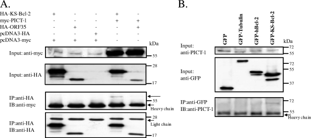
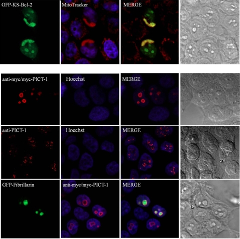
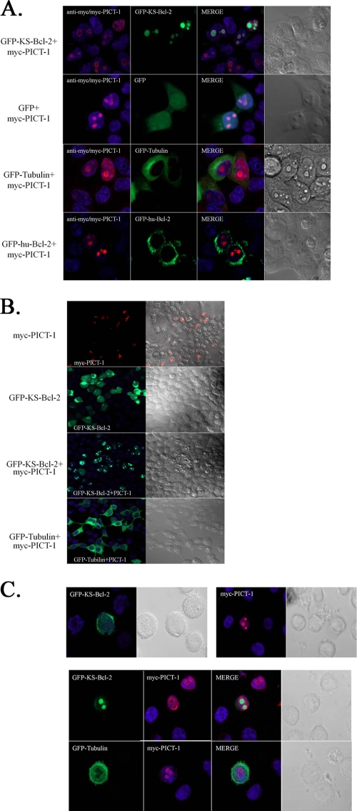
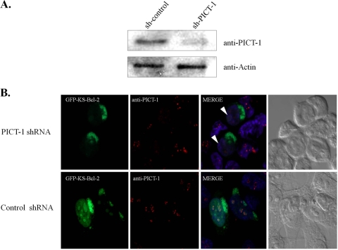
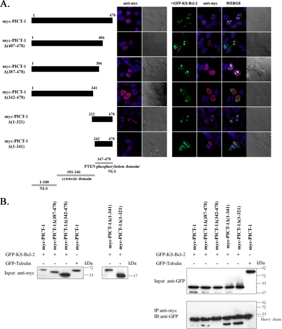
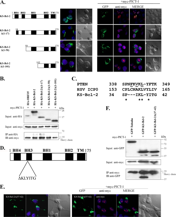
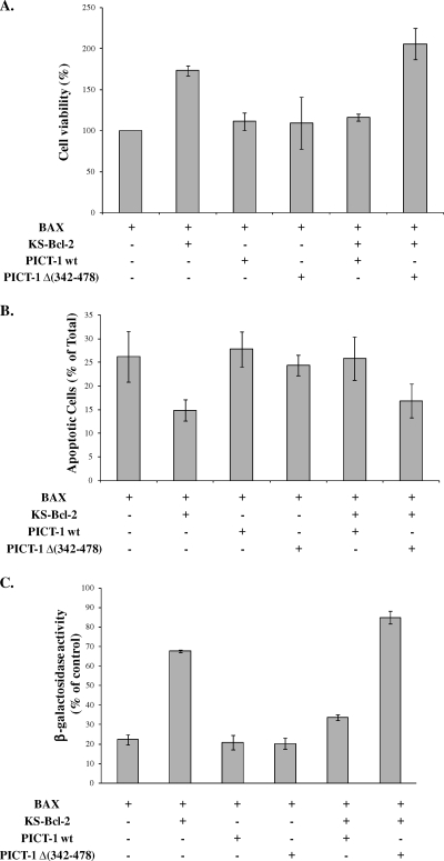
KS-Bcl-2, encoded by Kaposi's sarcoma-associated herpesvirus (KSHV), is a structural and functional homologue of the Bcl-2 family of apoptosis regulators. Like several other Bcl-2 family members, KS-Bcl-2 protects cells from apoptosis and autophagy. Using a yeast two-hybrid screen and coimmunoprecipitation assays, we identified a novel KS-Bcl-2-interacting protein, referred to as protein interacting with carboxyl terminus 1 (PICT-1), encoded by a candidate tumor suppressor gene, GLTSCR2. Confocal laser scanning microscopy revealed nucleolar localization of PICT-1, whereas KS-Bcl-2 was located mostly at the mitochondrial membranes with a small fraction in the nucleoli. Ectopic expression of PICT-1 resulted in a large increase in the nucleolar fraction of KS-Bcl-2, and only a minor fraction remained in the cytoplasm. Furthermore, knockdown of endogenous PICT-1 abolished the nucleolar localization of KS-Bcl-2. However, ectopically expressed PICT-1 did not alter the cellular distribution of human Bcl-2. Subsequent analysis mapped the crucial amino acid sequences of both KS-Bcl-2 and PICT-1 required for their interaction and for KS-Bcl-2 targeting to the nucleolus. Functional studies suggest a correlation between nucleolar targeting of KS-Bcl-2 by PICT-1 and reduction of the antiapoptotic activity of KS-Bcl-2. Thus, these studies demonstrate a cellular mechanism to sequester KS-Bcl-2 from the mitochondria and to downregulate its virally encoded antiapoptotic activity. Additional characterization of the interaction of KS-Bcl-2 and PICT-1 is likely to shed light on the functions of both proteins.


Figures

FIG. 1.
FIG. 1.
KS-Bcl-2 physically associates with PICT-1 in 293T cells. (A) To detect the association between PICT-1 and KS-Bcl-2, expression vectors containing HA-KS-Bcl-2 and myc-PICT-1 were transfected into 293T cells. Cells transfected with the HA-KS-Bcl-2 expression vector, together with control empty vector (pcDNA-myc), were used as negative controls. myc-PICT-1 cotransfected with HA-tagged ORF35 (HA-ORF35) served as an additional control. After 24 h, whole-cell extracts were prepared and 30-μg aliquots were analyzed for protein expression with antibodies to HA and myc (Input). Next, 400 μg of the lysates was subjected to immunoprecipitation (IP) with anti-HA, followed by sequential Western blotting (IB) with anti-myc and anti-HA antibodies. As indicated by the arrows, myc-PICT-1 coprecipitated with HA-KS-Bcl-2 but not with the unrelated protein HA-ORF35. (B) Similarly, GFP, GFP-tubulin, GFP-hBcl-2, and GFP-KS-Bcl-2 expression vectors were transfected into 293T cells. After 24 h, whole-cell extracts were prepared and analyzed for protein expression with antibodies to PICT-1 and GFP to detect the endogenous and exogenous proteins, respectively (Input). Whole-cell lysates were then immunoprecipitated with anti-GFP and immunoblotted with anti-PICT-1 antibody.
FIG. 2.
FIG. 2.
Cellular localization of KS-Bcl-2 and PICT-1. 293T cells were transfected with the indicated expression plasmids. Mitochondrial localization of KS-Bcl-2 was confirmed with MitoTracker. Ectopically expressed myc-PICT-1 was detected by anti-myc, while endogenous expression was identified with anti-PICT-1 antibody. GFP-fibrillarin was used as a marker for nucleoli. The corresponding staining of nuclear DNA by Hoechst and differential interference contrast (DIC) imaging is also displayed.
FIG. 3.
FIG. 3.
KS-Bcl-2 accumulates in the nucleoli when coexpressed with PICT-1. (A) 293T cells were transfected with the indicated expression plasmids. Overexpression of PICT-1 increased the nucleolar localization of KS-Bcl-2 but failed to affect the cellular localization of GFP, GFP-tubulin, or GFP-hu-Bcl-2. Hoechst staining was used to visualize cell nuclei, and DIC images were used to demonstrate the morphology of the cells. (B) Lower magnification showing the extensive and selective effect of the expression of PICT-1 on the cellular localization of GFP-KS-Bcl-2 in transfected 293T cells. (C) BCBL-1 cells were transfected with the indicated expression plasmids and visualized as described above.
FIG. 4.
FIG. 4.
Knockdown of endogenous PICT-1 aborogates the nucleolar localization of KS-Bcl-2. (A) pSilencer 3.1 H1-hygro plasmids encoding shRNA targeting PICT-1 or control shRNA were transfected into 293T cells, and 24 h after transfection, 100 μg/ml hygomycin was added. After 4 days, a GFP-KS-Bcl-2 expression vector was transfected, and protein cell extracts were prepared and assayed with antibodies to PICT-1 to confirm knockdown; anti-actin was included as a loading control. (B) In parallel, cells were fixed, and slides were prepared and viewed as described for panel A. The arrowheads indicate cells in which the expression of PICT-1 was knocked down and KS-Bcl-2 was not found the nucleoli.
FIG. 5.
FIG. 5.
Mapping the nucleolar localization signal of PICT-1 and the requirements for KS-Bcl-2 interaction. (A) Schematic representations of wild-type PICT-1 and its deletion mutants used in the mapping experiments; the boundary amino acids are numbered. The results of previously published functional mapping of PICT-1 (40) are shown in gray. 293T cells were transfected with the indicated myc-PICT-1 expression plasmid alone or with GFP-KS-Bcl-2, and the subcellular localization of the respective deletion mutant proteins was determined by immunofluorescence after staining them with anti-myc antibody and Hoechst. (B) Western blots showing coprecipitation of full-length and truncated myc-PICT-1 and KS-Bcl-2. Extracts of 293T cells cotransfected with plasmids encoding KS-Bcl-2 and full-length myc-PICT-1 or the indicated PICT-1 deletion mutants were first examined for exogenous protein expression with anti-GFP and anti-myc antibodies. The expression of full-length and truncated myc-PICT-1 is presented in two separate gels of 12% and 15% PAGE displaying their size differences (Input). Whole-cell extracts were immunoprecipitated with anti-myc antibody, and the presence of KS-Bcl-2 in the precipitates was examined by probing with anti-GFP antibody. GFP-tubulin was used as a negative control.
FIG. 6.
FIG. 6.
Mapping the binding domain of KS-Bcl-2 to PICT-1. (A) Schematic representations of wild-type KS-Bcl-2 and its deletion mutants used in the mapping experiments; the boundary amino acids are numbered. The locations of the BH-1 to -4 motifs and the transmembrane domain are indicated. 293T cells were transfected with the indicated GFP-KS-Bcl-2 expression plasmid alone or with myc-PICT-1, and the subcellular localization of the respective deletion mutant proteins was determined by immunofluorescence after staining them with anti-myc antibody and Hoechst. (B) Western blots showing coprecipitation of KS-Bcl-2 mutants and PICT-1. Extracts of 293T cells cotransfected with plasmids encoding PICT-1 and full-length KS-Bcl-2 or the indicated KS-Bcl-2 deletion mutants were first examined for exogenous protein expression with anti-HA and anti-myc antibodies (Input). Whole-cell extracts were then immunoprecipitated with anti-HA antibody, and the presence of PICT-1 in the precipitates was examined by probing with anti-myc antibody. (C) Alignment of potential PICT-1-binding motifs of PTEN, ICP0, and KS-Bcl-2. PTEN mutations that lost their binding to PICT-1 are underlined. (D and E) Schematic diagram of KS-Bcl-2Δ(37-42) (D) and its cellular localization when expressed alone or together with myc-PICT-1 (E). (F) Plasmids encoding myc-PICT-1 were cotransfected with GFP-tubulin, GFP-KS-Bcl-2, or GFP-KS-Bcl-2Δ(37-42) mutant into 293T cells, and exogenous protein expression was verified (Input). Whole-cell extracts were then immunoprecipitated with anti-myc antibody, and the presence of KS-Bcl-2 in the precipitates was examined by probing with anti-GFP antibody.
FIG. 7.
FIG. 7.
PICT-1 reduces the antiapoptotic activity of KS-Bcl-2. (A) 293T cells were transfected with the indicated plasmids, together with GFP expression plasmid to mark the transfected cells. All samples received equal amounts of DNA. The viability of the cells was determined at 24 h posttransfection by scoring the percentage of transfected cells that were alive and/or nonapoptotic (>130 GFP-positive cells in 15 independent fields were counted blindly per sample). Means ± standard deviations of three independent experiments are shown. (B) 293T cells were cotransfected with the indicated expression vectors. Apoptosis was measured at 20 h posttransfection via flow cytometry by staining the cells with annexin V-FITC and PI. The data are presented as percentages of annexin V-positive cells and represent the means ± standard deviations of five experiments. (C) 293T cells were cotransfected with the indicated expression vectors, together with a vector for β-Gal. Cells were harvested 24 h posttransfection and assayed for β-Gal activity. The results shown are means ± standard deviations (n = 3) and are representative of three independent experiments. wt, wild type.

Similar articles

Cited by

References

    1. Andersen, J. S., Y. W. Lam, A. K. Leung, S. E. Ong, C. E. Lyon, A. I. Lamond, and M. Mann. 2005. Nucleolar proteome dynamics. Nature 433:77-83. - PubMed
    1. Barboule, N., I. Truchet, and A. Valette. 2005. Localization of phosphorylated forms of Bcl-2 in mitosis: co-localization with Ki-67 and nucleolin in nuclear structures and on mitotic chromosomes. Cell Cycle 4:590-596. - PubMed
    1. Bellows, D. S., B. N. Chau, P. Lee, Y. Lazebnik, W. H. Burns, and J. M. Hardwick. 2000. Antiapoptotic herpesvirus Bcl-2 homologs escape caspase-mediated conversion to proapoptotic proteins. J. Virol. 74:5024-5031. - PMC - PubMed
    1. Bellows, D. S., M. Howell, C. Pearson, S. A. Hazlewood, and J. M. Hardwick. 2002. Epstein-Barr virus BALF1 is a BCL-2-like antagonist of the herpesvirus antiapoptotic BCL-2 proteins. J. Virol. 76:2469-2479. - PMC - PubMed
    1. Boisvert, F. M., S. van Koningsbruggen, J. Navascues, and A. I. Lamond. 2007. The multifunctional nucleolus. Nat. Rev. Mol. Cell Biol. 8:574-585. - PubMed

Publication types

MeSH terms