. 2010 Mar;84(6):2963-71.
doi: 10.1128/JVI.02015-09. Epub 2009 Dec 30.

Interaction domains of the UL16 and UL21 tegument proteins of herpes simplex virus

Affiliations

Interaction domains of the UL16 and UL21 tegument proteins of herpes simplex virus

Amy L Harper et al. J Virol. 2010 Mar.

Abstract

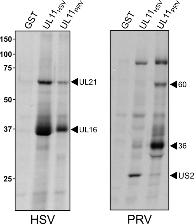
The UL16 protein of herpes simplex virus is capsid associated and was previously identified as a binding partner of the membrane-associated UL11 tegument protein (J. S. Loomis, R. J. Courtney, and J. W. Wills, J. Virol. 77:11417-11424, 2003). In those studies, a less-prominent, approximately 65-kDa binding partner of unknown identity was also observed. Mass spectrometry studies have now revealed this species to be UL21, a tegument protein that has been implicated in the transport of capsids in the cytoplasm. The validity of the mass spectrometry results was tested in a variety of coimmunoprecipitation and glutathione S-transferase pull-down experiments. The data revealed that UL21 and UL16 can form a complex in the absence of other viral proteins, even when the assays used proteins purified from Escherichia coli. Moreover, UL11 was able to pull down UL21 only when UL16 was present, suggesting that all three proteins can form a complex. Deletion analyses revealed that the second half of UL21 (residues 268 to 535) is sufficient for the UL16 interaction and packaging into virions; however, attempts to map a subdomain of UL16 were largely unsuccessful, with only the first 40 (of 373) residues being found to be dispensable. Nevertheless, it is clear that UL16 must have two distinct binding sites, because covalent modification of its free cysteines with N-ethylmaleimide blocked binding to UL11 but not UL21. These findings should prove useful for elucidating the molecular machinery used to transmit a signal into a virion when it attaches to cells, a recently discovered mechanism in which UL16 is a central player.


Figures

FIG. 1.
FIG. 1.
Distinct interaction profiles observed with GST-UL11 homologs of HSV and PRV. Vero cells were infected with HSV or PRV, and 5 h later, newly synthesized proteins were radiolabeled with [35S]methionine-cysteine for 3 h. The cells were lysed with NP-40-containing buffer, and after discarding the nuclei, glutathione beads that displayed the indicated GST-fusion proteins were added. Bound proteins were separated by SDS-PAGE, transferred to nitrocellulose, and detected by autoradiography.
FIG. 2.
FIG. 2.
Coimmunoprecipitation of UL16 and UL21 from infected cells. Vero cells infected with HSV (A) or HSV.UL16-CFP (B) were disrupted in NP-40 lysis buffer 20 h postinfection. Proteins were immunoprecipitated (IP) with antibodies specific for UL11, UL16, UL21, or GFP or no herpesvirus proteins (preimmune [pre]). The proteins were separated by SDS-PAGE and analyzed by immunoblotting with anti-UL16 or anti-UL21 serum. A portion (1/15) of the lysate was analyzed for protein expression.
FIG. 3.
FIG. 3.
GST-UL11 pulls down UL21 only when UL16 is present. Vero cells infected for 5 h with wild-type HSV or HSV.ΔUL16 were radiolabeled with [35S]methionine-cysteine for 3 h, washed, and resuspended in NP-40 lysis buffer. The indicated GST constructs were added, and bound proteins were separated by SDS-PAGE, transferred to nitrocellulose, and subjected to autoradiography (left panel). The blot was stained with Ponceau S to visualize the amounts of purified GST proteins present (right panel).
FIG. 4.
FIG. 4.
The UL16-binding site is located in the second half of UL21. (A) Diagrams of UL21 and the N- and C-terminal truncation mutants, all of which were constructed as GFP-fusion proteins. A summary of all the binding results is provided on the right. (B) Expression levels of GFP-UL21 derivatives. Vero cells were transfected with the indicated constructs, and 20 h later, they were lysed in sample buffer, separated by SDS-PAGE, and transferred to nitrocellulose for immunoblotting with antibodies specific for GFP. (C) GST-UL16 pull-down assays. Vero cells transfected with the indicated constructs were harvested in NP-40-containing lysis buffer, and after discarding the nuclei, glutathione beads that displayed the GST-UL16 fusion protein were added. Bound proteins were separated by SDS-PAGE, transferred to nitrocellulose, and detected with GFP-specific antibodies.
FIG. 5.
FIG. 5.
Nuclear localization of GFP-UL21 in the absence of other viral proteins. Confocal microscopy results for GFP-tagged UL21 constructs are shown. Vero cells were seeded on coverslips and subsequently transfected with the indicated GFP-UL21 constructs. After 20 h, the cells were stained with Hoechst dye to label the nuclei, washed, paraformaldehyde fixed, affixed to slides, and examined with a confocal microscope to visualize the GFP signals alone (left panels) or in combination with the nuclear signals (right panels). For the bottom panels (below the dotted line), cells that had been transfected for 18 h were infected with HSV and examined at the indicated times postinfection (PI).
FIG. 6.
FIG. 6.
The second half of UL21 is sufficient for packaging into virions. Vero cells were transfected to express the indicated GFP-UL21 constructs or GFP alone and then infected with HSV at 24 h posttransfection. After another 20 h, extracellular virions were purified from the medium by centrifugation through a sucrose cushion. Virions and infected cell lysates were separated by SDS-PAGE and analyzed by immunoblotting with antibodies specific for GFP and VP5.
FIG. 7.
FIG. 7.
UL21 and UL16 directly interact, but this is not essential for UL21 packaging. The indicated GST-fusion proteins were expressed in bacteria and purified on glutathione-Sepharose beads. These were incubated with increasing amounts of soluble His6-UL16 (100, 200, or 400 ng), which was also purified from bacteria. The beads were washed, and the associated proteins were separated by SDS-PAGE and then transferred to nitrocellulose. (A) The amounts of input His6-UL16 and each of the GST-fusion proteins were revealed by Ponceau S staining. (B) The amount of His6-UL16 bound to the beads was measured by immunoblotting with a rabbit antibody that recognizes the His6 tag. (C) Media from Vero cells infected with either wild-type or ΔUL16 HSV were collected 24 h postinfection and centrifuged to pellet the virions. Samples of the cell lysates and virions were separated by SDS-PAGE and analyzed by immunoblotting with the indicated antibodies.
FIG. 8.
FIG. 8.
NEM modification of UL16 blocks the interaction with UL11 but not UL21. Insect cells infected with a baculovirus that expresses UL16-GFP were lysed with NP-40 buffer and either untreated (lane 1 of each panel) or treated for 30 min with NEM, either before or after 2 h of incubation with the indicated bead-bound GST-fusion protein (lanes 2 and 4 of each panel). Alternatively, NEM was added to the GST-fusion protein and washed away prior to mixing with UL16-GFP (lane 3 in each panel). The bead-bound GST fusion and associated proteins were washed, dissolved in sample buffer, separated in SDS-10% polyacrylamide gels, and visualized by immunoblotting with anti-GST (bottom) and anti-GFP (top) antibodies.

Similar articles

Cited by

References

    1. Addison, C., F. J. Rixon, and V. G. Preston. 1990. Herpes simplex virus type 1 UL28 gene product is important for the formation of mature capsids. J. Gen. Virol. 71:2377-2384. - PubMed
    1. Baines, J. D., C. Cunningham, D. Nalwanga, and A. Davison. 1997. The UL15 gene of herpes simplex virus type 1 contains within its second exon a novel open reading frame that is translated in frame with the UL15 gene product. J. Virol. 71:2666-2673. - PMC - PubMed
    1. Baines, J. D., A. H. Koyama, T. Huang, and B. Roizman. 1994. The UL21 gene products of herpes simplex virus 1 are dispensable for growth in cultured cells. J. Virol. 68:2929-2936. - PMC - PubMed
    1. Baines, J. D., and B. Roizman. 1991. The open reading frames UL3, UL4, UL10, and UL16 are dispensable for the replication of herpes simplex virus 1 in cell culture. J. Virol. 65:938-944. - PMC - PubMed
    1. Baines, J. D., and B. Roizman. 1992. The UL11 gene of herpes simplex virus 1 encodes a function that facilitates nucleocapsid envelopment and egress from cells. J. Virol. 66:5168-5174. - PMC - PubMed

Publication types

MeSH terms

Substances

LinkOut - more resources