. 2010 Mar;84(6):3059-67.
doi: 10.1128/JVI.02044-09. Epub 2009 Dec 30.

Serotype-specific structural differences in the protease-cofactor complexes of the dengue virus family

Affiliations

Serotype-specific structural differences in the protease-cofactor complexes of the dengue virus family

Sumana Chandramouli et al. J Virol. 2010 Mar.

Abstract

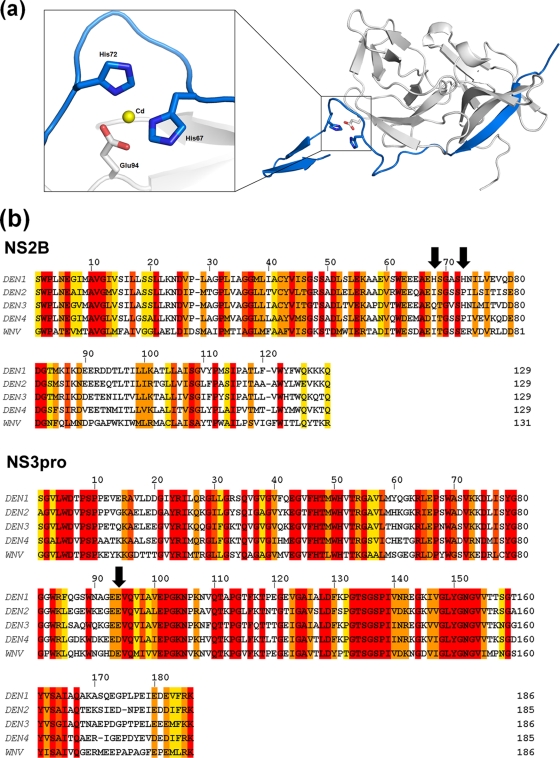
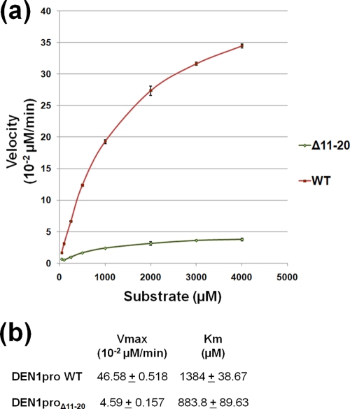
With an estimated 40% of the world population at risk, dengue poses a significant threat to human health, especially in tropical and subtropical regions. Preventative and curative efforts, such as vaccine development and drug discovery, face additional challenges due to the occurrence of four antigenically distinct serotypes of the causative dengue virus (DEN1 to -4). Complex immune responses resulting from repeat assaults by the different serotypes necessitate simultaneous targeting of all forms of the virus. One of the promising targets for drug development is the highly conserved two-component viral protease NS2B-NS3, which plays an essential role in viral replication by processing the viral precursor polyprotein into functional proteins. In this paper, we report the 2.1-A crystal structure of the DEN1 NS2B hydrophilic core (residues 49 to 95) in complex with the NS3 protease domain (residues 1 to 186) carrying an internal deletion in the N terminus (residues 11 to 20). While the overall folds within the protease core are similar to those of DEN2 and DEN4 proteases, the conformation of the cofactor NS2B is dramatically different from those of other flaviviral apoprotease structures. The differences are especially apparent within its C-terminal region, implicated in substrate binding. The structure reveals for the first time serotype-specific structural elements in the dengue virus family, with the reported alternate conformation resulting from a unique metal-binding site within the DEN1 sequence. We also report the identification of a 10-residue stretch within NS3pro that separates the substrate-binding function from the catalytic turnover rate of the enzyme. Implications for broad-spectrum drug discovery are discussed.


Figures

FIG. 1.
FIG. 1.
Thermostability assays to identify the optimal buffer and salt for DEN1pro. (a) Increase in fluorescence upon binding of Sypro-Orange dye to the DEN1pro protein when subjected to thermal ramping from 20°C to 95°C in the presence of various buffers. A pH range of 4 to 10.5 was tested, with a representative set of 12 buffers out of a total 32 tested shown here. (b) Negative-derivative plot (−dF/dT) of panel a, with the temperature at the point of inflection giving the Tm of the protein. (c) Effects of various salts on the Tm of DEN1pro in the presence of the optimal buffer identified in panel b, sodium cacodylate, pH 6.5. Shown is a negative-derivative plot of a fluorescence assay similar to that in panel a, with 10 out of 24 salts tested shown for clarity.
FIG. 2.
FIG. 2.
Structure of DEN1proΔ11-20. (a) Structures of DEN1proΔ11-20 (left) and its active-site S135A mutant (right). The NS3pro core is shown in green, and the NS2B cofactor is shown in blue. (b) Superimposition of the DEN1proΔ11-20 NS3 core over DEN2pro NS3 (gold; PDB code, 2FOM) and DEN4pro (magenta; PDB code, 2VBC) showing overall similar folds. L18 and Q167 mark the N and C termini of DEN2pro. (c) Enlarged view of the boxed region in panel b showing the shift in the β hairpins between DEN1proΔ11-20 and DEN2pro (left) and the C-terminal tail of the three DENV proteases (right). DEN1proΔ11-20, green; DEN2pro, gold; DEN4pro, magenta.
FIG. 3.
FIG. 3.
Conformation of the DEN1proΔ11-20 active site. (a) Change in conformation of Asn152 in the presence of a free hydroxyl group on Ser135 (left) compared to the S135A mutant (right). The essential hydrogen bond between the active-site His51 and Ser135 is disrupted by the presence of a Ni2+ atom (cyan) coordinated with His51 and a water molecule (gray). (b) Conformation of Asn152 in other flaviviral proteases, DEN2pro (PDB code, 2FOM) and WNVpro H51A (PDB code, 2GGV).
FIG. 4.
FIG. 4.
Conformation of DEN1proΔ11-20 NS2Bc. Shown is NS2Bc (blue) of DEN1proΔ11-20 (a), DEN2pro (b), and WNVpro (c) in complex with a peptidic inhibitor. The positions of Trp61 and Ser68 and the corresponding residues in WNVpro, Trp62, and Thr69 are shown as sticks. The corresponding NS3pro structures are represented as ribbons (gray).
FIG. 5.
FIG. 5.
A unique metal-coordinated site in DEN1proΔ11-20. (a) A metal-coordinated site composed of His67 and His72 of NS2Bc (blue) with Glu94 (gray) of NS3. The central cadmium atom is represented in yellow. (b) Sequence alignment of the NS2B and NS3pro sequences of DEN1 to -4 and WNV. The arrows indicate His67, His72, and Glu94 of DEN1. The alignment was generated using ClustalW2 (European Bioinformatics Institute [http://www.ebi.ac.uk/]). The shaded residues are colored according to the degree of conservation, ranging from yellow (least) to red (most conserved).
FIG. 6.
FIG. 6.
Enzyme kinetics of DEN1proΔ11-20. (a) Enzyme reaction kinetics for DEN1proΔ11-20 (green) compared to WT DEN1pro (red). The reactions were carried out with the peptide substrate FAAGRK-pNA, corresponding to the NS3/4A junction. Free pNA was measured as absorbance at 405 nm and converted to the concentration released using a pNA standard curve. At least triplicates of reactions, with substrate concentrations ranging from 50 μM to 4 mM and the enzyme at 0.4 μM, were tested. (b) The Vmax and Km for WT DEN1pro and DEN1proΔ11-20 were calculated based on Michaelis-Menten kinetics using GraphPad Prism software.

References

    1. Adams, P. D., R. W. Grosse-Kunstleve, L. W. Hung, T. R. Ioerger, A. J. McCoy, N. W. Moriarty, R. J. Read, J. C. Sacchettini, N. K. Sauter, and T. C. Terwilliger. 2002. PHENIX: building new software for automated crystallographic structure determination. Acta Crystallogr. D Biol. Crystallogr. 58:1948-1954. - PubMed
    1. Aleshin, A. E., S. A. Shiryaev, A. Y. Strongin, and R. C. Liddington. 2007. Structural evidence for regulation and specificity of flaviviral proteases and evolution of the Flaviviridae fold. Protein Sci. 16:795-806. - PMC - PubMed
    1. Collaborative Computational Project Number 4. 1994. The CCP4 suite: programs for protein crystallography. Acta Crystallogr. D Biol. Crystallogr. 50:760-763. - PubMed
    1. De Clercq, E. 2007. The design of drugs for HIV and HCV. Nat. Rev. Drug Discov. 6:1001-1018. - PubMed
    1. Ekonomiuk, D., X. C. Su, K. Ozawa, C. Bodenreider, S. P. Lim, Z. Yin, T. H. Keller, D. Beer, V. Patel, G. Otting, A. Caflisch, and D. Huang. 2009. Discovery of a non-peptidic inhibitor of West Nile virus NS3 protease by high-throughput docking. PLoS Negl. Trop. Dis. 3:e356. - PMC - PubMed

Publication types

MeSH terms

LinkOut - more resources