. 2010 Mar;84(6):2753-61.
doi: 10.1128/JVI.01813-09. Epub 2009 Dec 30.

Canine distemper viruses expressing a hemagglutinin without N-glycans lose virulence but retain immunosuppression

Affiliations

Canine distemper viruses expressing a hemagglutinin without N-glycans lose virulence but retain immunosuppression

Bevan Sawatsky et al. J Virol. 2010 Mar.

Abstract

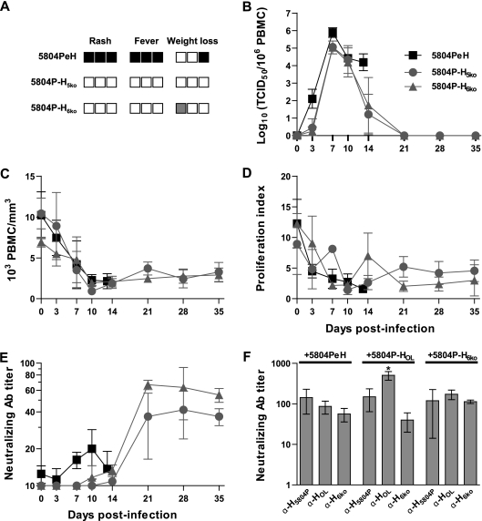
Paramyxovirus glycoproteins are posttranslationally modified by the addition of N-linked glycans, which are often necessary for correct folding, processing, and cell surface expression. To establish the contribution of N glycosylation to morbillivirus attachment (H) protein function and overall virulence, we first determined the use of the potential N-glycosylation sites in the canine distemper virus (CDV) H proteins. Biochemical characterization revealed that the three sites conserved in all strains were N glycosylated, whereas only two of the up to five additional sites present in wild-type strains are used. A wild-type virus with an H protein reproducing the vaccine strain N-glycosylation pattern remained lethal in ferrets but with a prolonged course of disease. In contrast, introduction of the vaccine H protein in the wild-type context resulted in complete attenuation. To further characterize the role of N glycosylation in CDV pathogenesis, the N-glycosylation sites of wild-type H proteins were successively deleted, including a nonstandard site, to ultimately generate a nonglycosylated H protein. Despite reduced expression levels, this protein remained fully functional. Recombinant viruses expressing N-glycan-deficient H proteins no longer caused disease, even though their immunosuppressive capacities were retained, indicating that reduced N glycosylation contributes to attenuation without affecting immunosuppression.


Figures

FIG. 1.
FIG. 1.
Construction and characterization of recombinant viruses with vaccine-like H protein N glycosylation. (A) Schematic drawing of the H5804P and HOL proteins. The HOL-like protein has the primary sequence of H5804P with the mutations N309Q, N391Q, N456Q, and S605stop to reproduce the glycosylation pattern of HOL. Protein schematics are shown from the N to the C terminus. The N-terminal box represents the cytoplasmic tail, the black box represents the transmembrane domain, and the C-terminal box represents the C-terminal ectodomain. (B) Western blot analysis of vaccine-like H protein expression. H5804P, HOL, and HOL-like were expressed in Vero dogSLAMtag cells by transfection of expression plasmids and detected by Western blotting. Each lysate was also treated with PNGase F to remove all N-glycans. (C) Cell surface biotinylation of vaccine-like H proteins. Cells transfected with expression plasmids for H5804P, HOL, or HOL-like were labeled with Sulfo-NHS-LC-biotin, immunoprecipitated (IP) with an antiserum recognizing the H protein cytoplasmic tail, and detected with a streptavidin-HRP conjugate. Western blots of cell lysates from the same experiment were analyzed for total H protein expression. The relative surface expression of each protein was calculated by normalizing the biotinylation or Western blot signal to the respective H5804P signal. The relative surface expression represents the ratio between the normalized biotinylation signal and the normalized Western blot signal for each replicate. Average relative surface expression is shown, and the blots are representative of three independent replicates. IB, immunoblot. (D) Cell-cell fusion observed for wild-type and mutant H proteins. Confluent monolayers of Vero dogSLAMtag cells were transfected with F5804P in combination with H5804P, HOL, or HOL-like expression plasmid. The images were taken 24 h posttransfection at ×100 magnification. The left image shows control untransfected Vero dogSLAMtag cells. (E and F) Growth kinetics of recombinant 5804P viruses expressing either H5804P, HOL, or HOL-like in Vero dogSLAMtag cells shown as either cell-associated (E) or released (F) virus. The cells were infected at an MOI of 0.01, and samples were harvested daily for 5 days. Titers are expressed as TCID50/ml, and the error bars indicate standard deviations.
FIG. 2.
FIG. 2.
Virulence of vaccine-like viruses in ferrets. Groups of three to six animals were infected intranasally with 105 TCID50 of either the recombinant parental 5804PeH virus or the recombinant viruses 5804P-HOL and 5804P-HOL-like. (A) Virulence index. Each box represents one animal, and black represents the highest (2), gray an intermediate (1), and white the lowest (0) score, as detailed in Materials and Methods. (B) Survival curve. Death of an animal is indicated by a step down on the curve. (C) The course of cell-associated viremia is shown as the log10 of the virus titer per 106 PBMCs. (D) CDV neutralizing-antibody response in plasma samples. Antibody titers are shown as reciprocals of the highest dilution in which CPE was observed. (E) Total leukocyte counts from infected animals, shown as 103 leukocytes per mm3. (F) In vitro proliferation activities of lymphocytes from infected animals. Days postinfection are indicated on the x axes of the graphs, and the error bars represent standard deviations.
FIG. 3.
FIG. 3.
Systematic deglycosylation of H5804P. (A) Schematic drawing of the N-glycosylation sites of the type N-X-S/T located in the ectodomain of H5804P and the mutant protein H5ko. Protein schematics are shown from the N terminus (left) to the C terminus (right). The N-terminal white boxes represent the cytoplasmic tail, the black boxes the transmembrane domain, and the C-terminal white boxes the ectodomain. (B) Western blot expression analysis of a comprehensive panel of N-glycosylation knockout mutant H5804P proteins. Proteins were expressed by transfection of expression plasmids in Vero dogSLAMtag cells. (Top) Single N-glycan knockout proteins (left blot) and the HD584N knock-in mutant protein (right blot) compared to H5804P protein. Note that HN309Q, HN603Q, and HD584N do not shift in apparent molecular weight. (Bottom) Quadruple- and quintuple-knockout proteins produced in all possible combinations. Mutant proteins are shown with H5804P and HN149,391,422Q to illustrate the observed molecular weight shift. The arrow indicates the higher-molecular-weight band observed for H5ko.
FIG. 4.
FIG. 4.
Identification of a nonstandard N-glycosylation site in H5804P and generation of recombinant viruses bearing N-glycan-deficient H proteins. (A) Schematic drawing of H5ko and the four remaining N-glycosylation sites located in the ectodomain, and H7ko, H9ko, and H6ko, indicating which potential N-glycosylation site is present in each protein. The site at position 152 is marked with an asterisk to indicate that it is the nonstandard type N-X-C. (B) Western blot comparing N-glycan-deficient H proteins to H5804P. Note that H9ko and H6ko comigrate with the lower-molecular-weight bands of H5ko and H7ko. The proteins were expressed by transfection of expression plasmids in Vero dogsSLAMtag cells. (C) Cell surface biotinylation of N-glycan-deficient H proteins. Cells transfected with expression plasmids for H5804P, H5ko, H7ko, or H6ko were labeled with Sulfo-NHS-LC-biotin, immunoprecipitated with an antiserum recognizing the H protein cytoplasmic tail, and detected with a streptavidin-HRP conjugate. Western blots of cell lysates from the same experiment were used to quantify total H protein expression. Average relative surface expression is shown, and the blots are representative of three independent replicates. (D) Cell-cell fusion observed for wild-type and mutant H proteins. Confluent monolayers of Vero dogSLAMtag cells were transfected with F5804P in combination with H5804P, H5ko, H7ko, or H6ko expression plasmid. The images were taken 24 h posttransfection at ×100 magnification. (E and F) Growth kinetics of the 5804PeH virus and the recombinant viruses expressing H5ko or H6ko in Vero dogSLAMtag cells shown as either cell-associated (E) or released (F) virus. The cells were infected at an MOI of 0.01, and samples were harvested daily for 5 days. Titers are expressed as TCID50/ml, and the error bars indicate standard deviations.
FIG. 5.
FIG. 5.
Virulence of N-glycan-deficient viruses in ferrets. Groups of three or four animals were infected intranasally with 105 TCID50 of either recombinant virus 5804P-H5ko or 5804P-H6ko. The results from the 5804PeH infections obtained in Fig. 2 are shown for comparison. (A) Virulence index. Each box represents one animal, and black represents the highest (2), gray an intermediate (1), and white the lowest (0) score, as detailed in Materials and Methods. (B) The course of cell-associated viremia is shown as the log10 of the virus titer per 106 PBMCs. (C) Total leukocyte counts, shown as 103 leukocytes per mm3. (D) In vitro proliferation activities of lymphocytes over the course of disease. (E) CDV neutralizing-antibody response in plasma samples. Antibody titers are shown as reciprocals of the highest dilution in which CPE was observed. Days postinfection are indicated on the x axes, and the error bars represent standard deviations. (F) Cross-neutralization of recombinant viruses. Plasma from animals infected with 5804PeH, 5804P-HOL, and 5804P-H6ko were tested for efficiency of neutralization against the respective viruses. Neutralizing-antibody titers are shown as reciprocals of the highest dilution at which CPE was observed. The asterisk indicates statistical significance for 5804P-HOL neutralization with α-HOL serum (P < 0.01) compared to neutralization with α-H5804P and α-H6ko sera, and the error bars represent standard deviations.

Similar articles

Cited by

References

    1. Anderson, D. E., and V. von Messling. 2008. The region between the canine distemper virus M and F genes modulates virulence by controlling fusion protein expression. J. Virol. 82:10510-10518. - PMC - PubMed
    1. Bagai, S., and R. A. Lamb. 1995. Individual roles of N-linked oligosaccharide chains in intracellular transport of the paramyxovirus SV5 fusion protein. Virology 209:250-256. - PubMed
    1. Baron, M. D. 2005. Wild-type Rinderpest virus uses SLAM (CD150) as its receptor. J. Gen. Virol. 86:1753-1757. - PubMed
    1. Bartz, R., U. Brinckmann, L. M. Dunster, B. Rima, V. ter Meulen, and J. Schneider-Schaulies. 1996. Mapping amino acids of measles virus hemagglutinin responsible for receptor (CD46) downregulation. Virology 224:334-337. - PubMed
    1. Bause, E., and G. Legler. 1981. The role of the hydroxy amino acid in the triplet sequence Asn-Xaa-Thr(Ser) for the N-glycosylation step during glycoprotein biosynthesis. Biochem. J. 195:639-644. - PMC - PubMed

Publication types

LinkOut - more resources