. 2010 Feb 1;184(3):1180-90.
doi: 10.4049/jimmunol.0901972. Epub 2009 Dec 30.

SHP-1 deficient mast cells are hyperresponsive to stimulation and critical in initiating allergic inflammation in the lung

Affiliations

SHP-1 deficient mast cells are hyperresponsive to stimulation and critical in initiating allergic inflammation in the lung

Li Zhang et al. J Immunol. .

Abstract

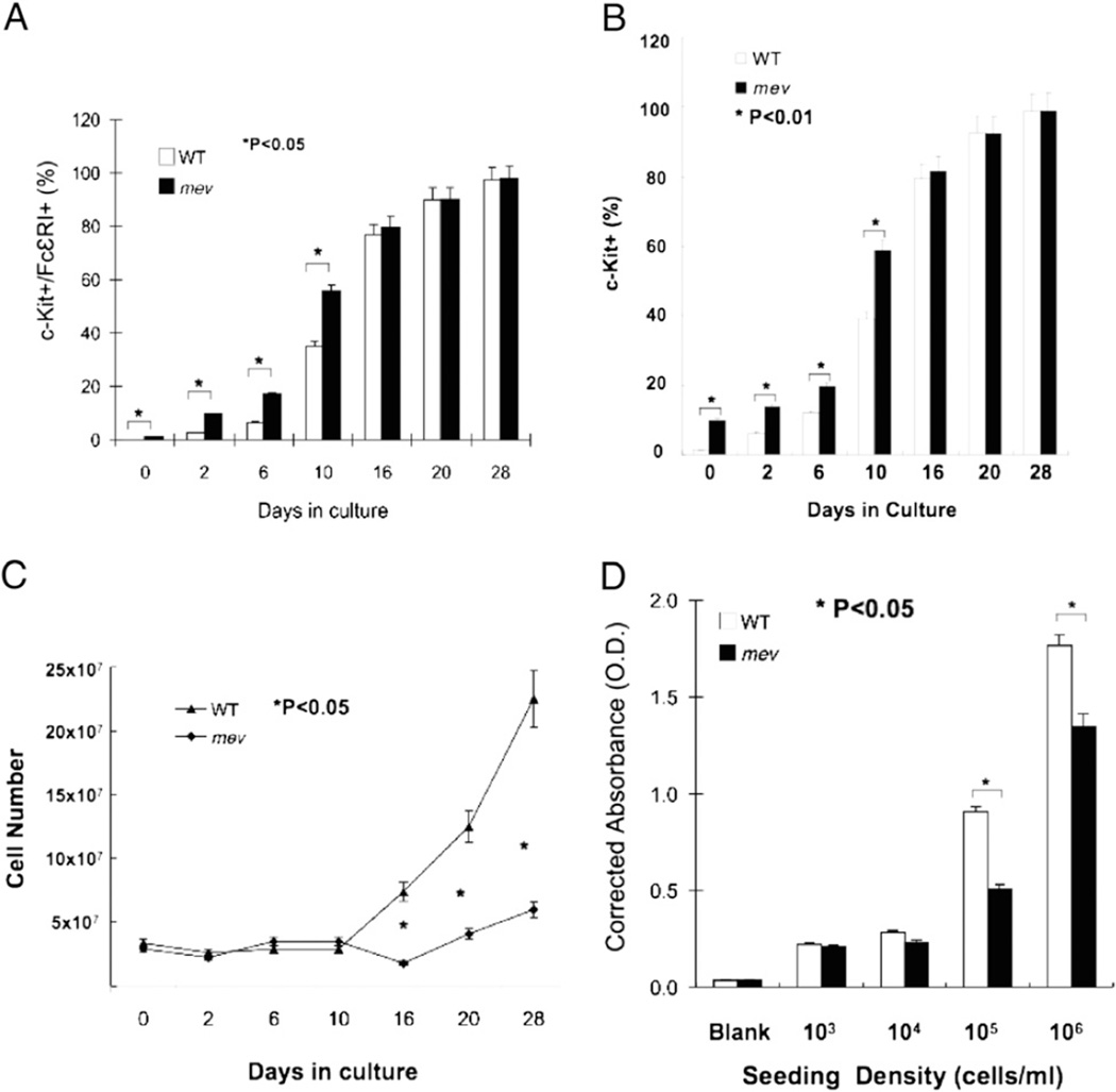
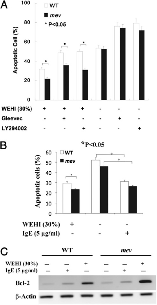
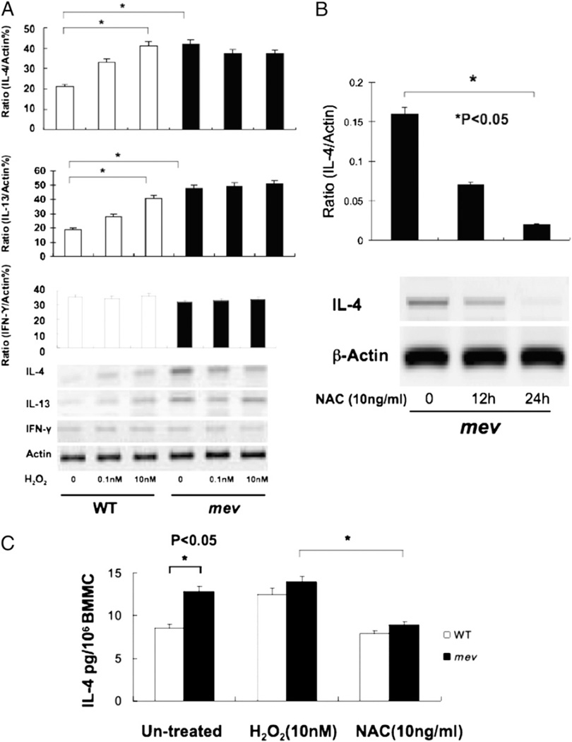
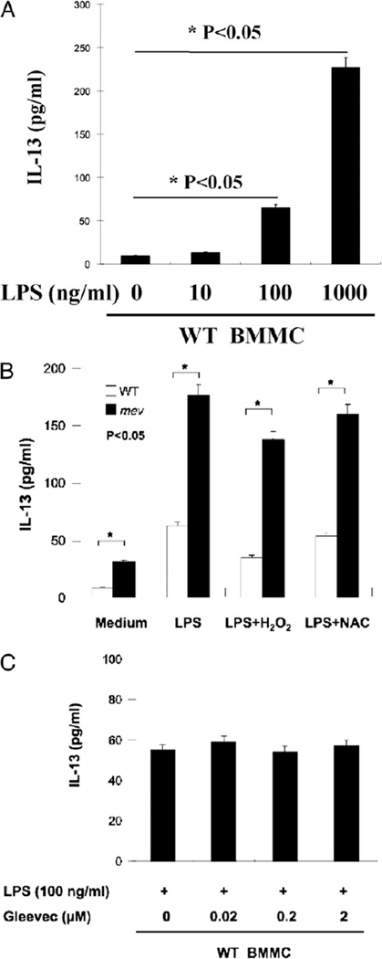
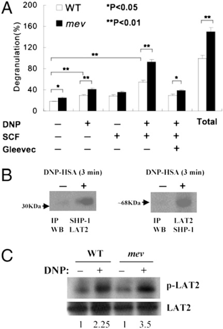
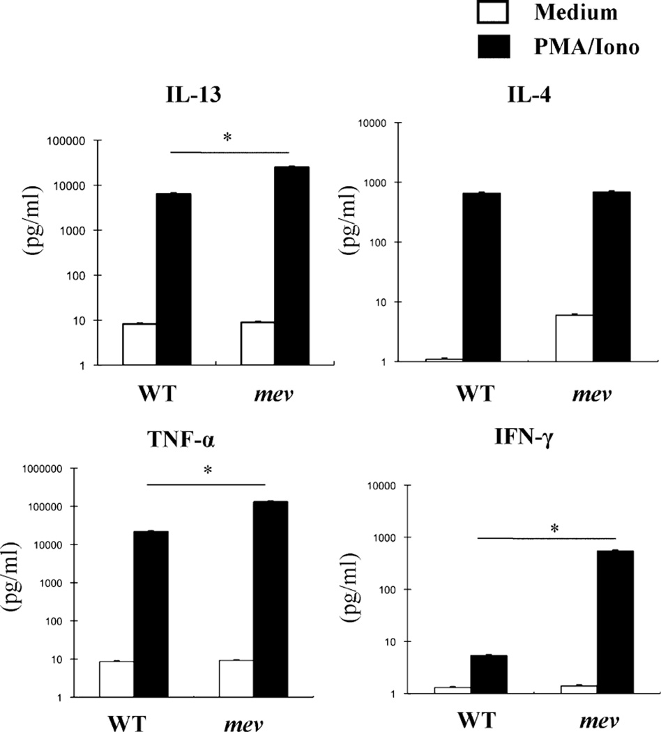
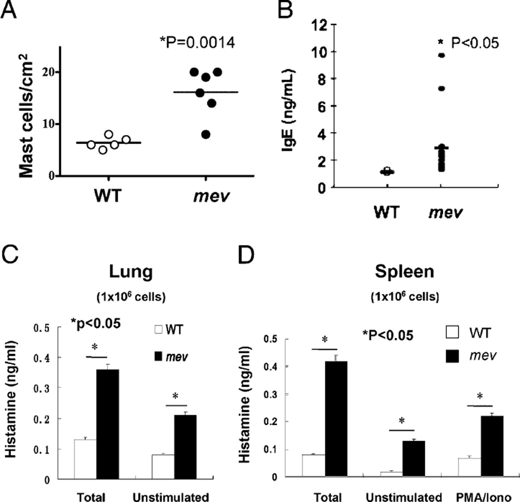
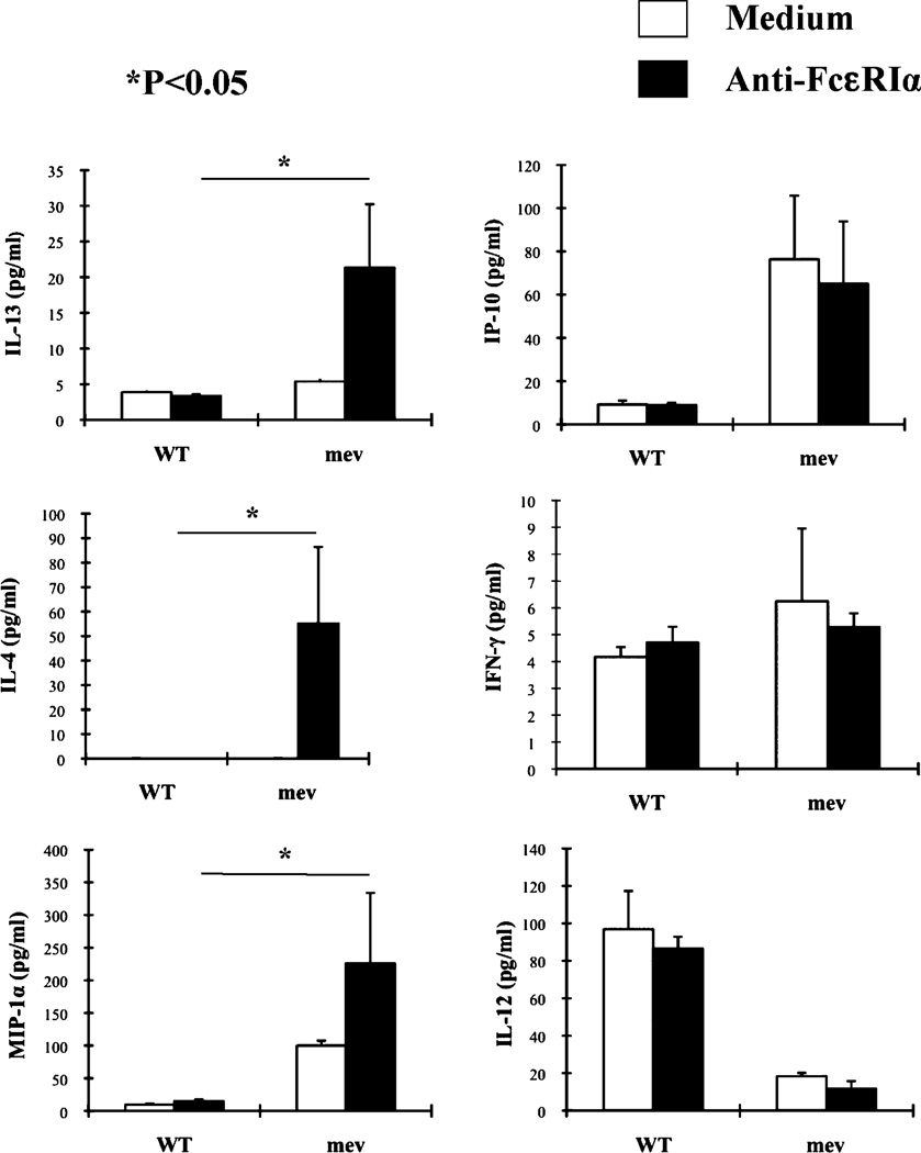
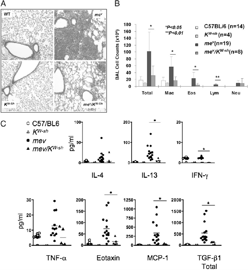
Phosphatase Src homology region 2 domain-containing phosphatase 1 (SHP-1)-deficient mice display an allergic asthma phenotype that is largely IL-13 and STAT6 dependent. The cell types responsible for the Th2 phenotype have not been identified. We hypothesized that SHP-1 deficiency leads to mast cell dysregulation and increased production and release of mediators and Th2 cytokines, leading to the allergic asthma phenotype. We examined SHP-1 regulation of mast cell differentiation, survival, and functional responses to stimulation using bone marrow-derived mast cells from viable motheaten (mev) mice. We assessed pulmonary phenotypical changes in mev mice on the mast cell-deficient Kit(W-Sh) genetic background. The results showed that SHP-1 deficiency led to increased differentiation and survival, but reduced proliferation, of mast cells. SHP-1-deficient mast cells produced and released increased amounts of mediators and Th2 cytokines IL-4 and -13 spontaneously and in response to H(2)O(2), LPS, and Fc epsilonI cross-linking, involving c-Kit-dependent and -independent processes. The Fc epsilonRI signaling led to binding of SHP-1 to linker for activation of T cells 2 and enhanced linker for activation of T cells 2 phosphorylation in mev bone marrow-derived mast cells. Furthermore, the number of mast cells in the lung tissue of mev mice was increased and mast cell production and release of Th2 cytokines were distinctly increased upon Fc epsilonRI stimulation. When backcrossed to the Kit(W-Sh) background, mev mice had markedly reduced pulmonary inflammation and Th2 cytokine production. These findings demonstrate that SHP-1 is a critical regulator of mast cell development and function and that SHP-1-deficient mast cells are able to produce increased Th2 cytokines and initiate allergic inflammatory responses in the lung.


Conflict of interest statement

Disclosures

The authors have no financial conflicts of interest.

Figures

FIGURE 1
FIGURE 1
BMMC differentiation and proliferation. At different time points, aliquots of cultured bone marrow cells from WT and mev mice were incubated with IgE overnight, stained for c-Kit and FcεRI, and analyzed by FACS. For each sample, 10,000 events were counted. Percentage of c-Kit/FcεRI double-positive cells (A) and percentage of c-Kit positive cells (B). Results are mean ± SD of eight pairs of WT and mev mice. C, Total number of bone marrow cells from each sample was determined before culture and at specified time points in culture. Results from three sets of independent experiments. D, After 4 wk in culture, mature BMMCs were serially diluted in conditioned medium and seeded in a microplate using 100 µl of the cells with indicated densities in triplicate. Medium alone was used as a baseline reference. Three days later, cell proliferation was determined by the XTT method. A representative of three experiments with similar results is shown.
FIGURE 2
FIGURE 2
Apoptosis of BMMCs under different conditions. After replacing WEHI-3B–conditioned medium, BMMCs were cultured in the presence or absence of WEHI-3B medium (30%), Gleevec (1 µM), or LY294002 (10 µM) for 24 h and analyzed by FACS with Annexin V and PI staining. A, The percentage of apoptotic cells. Three independent experiments were performed with triplicates for each sample. B, The percentage of apoptotic cells in the presence of IgE. C, RT-PCR analysis of expression of antiapoptosis gene Bcl-2 in WT and mev BMMCs. β-Actin mRNA was used as an internal control. A representative of two experiments is shown.
FIGURE 3
FIGURE 3
BMMC expression and secretion of IL-4 and -13 and IFN-γ in response to H2O2 stimulation. A, BMMCs (2 × 10⁶ cells/ml) were stimulated with indicated concentrations of H2O2 overnight, and cDNA from the cells was analyzed for IL-4 and -13 and IFN-γ mRNA expression. Densitometry analysis was performed on three independent experiments and normalized to β-actin (*p < 0.01). B, RT-PCR analysis of IL-4 expression after NAC treatment. cDNA from mev BMMCs treated with NAC (10 ng/ml) was analyzed for IL-4 expression at two time points. Densitometry was performed in three independent experiments and normalized to β-actin. C, IL-4 secretion by BMMCs after H2O2 (10 nM) stimulation or NAC (10 ng/ml) treatment. IL-4 protein in the BMMC culture medium was evaluated by ELISA after stimulation for 24 h (n = 8 for each group).
FIGURE 4
FIGURE 4
BMMC production of IL-13 in response to LPS. A, Dose-response of WT BMMCs to LPS stimulation. WT BMMCs (1 × 10⁷ cells/ml) were incubated with LPS of increasing concentrations for 24 h, and IL-13 in the supernatant was measured by ELISA (n = 3). B, IL-13 production by WT and mev BMMCs (1 × 10⁷ cells/ml) after incubation with LPS (100 ng/ml) with or without H2O2 or NAC (n = 5 for each group). The difference between mev BMMCs stimulated with LPS and LPS+H2O2 was not statistically significant (p > 0.05). C, Effect of Gleevec on LPS-stimulated BMMC production of IL-13. WT BMMCs were incubated with LPS for 24 h in the presence of varying concentrations of Gleevec, and IL-13 in the supernatant was measured by ELISA (n = 3 for each group).
FIGURE 5
FIGURE 5
BMMC degranulation in response to FcεRI and c-Kit signaling. A, BMMCs (1 × 10⁷ cells/ml) were sensitized with anti-DNP IgE overnight. Then cells were stimulated with DNP-HSA (100 ng), SCF (10 ng/ml), or a combination of DNP-HSA/SCF or DNP-HSA/SCF/Gleevec (1 µM). After 15 min, β-hexosaminidase released in the supernatant was measured by ELISA. Total amount of β-hexosaminidase was also determined after lysing unstimulated control cell samples. Data shown are mean percentage ± SD of triplicates of each sample relative to the total amount of β-hexosaminidase in WT control cells. Three independent experiments were performed with similar results. B, Binding of SHP-1 to LAT2. Protein samples from IgE-sensitized WT BMMCs stimulated with DNP-HSA or control were immunoprecipitated with anti–SHP-1 Ab and subsequently immunoblotted using anti-LAT2 Ab or vice versa. The relative positions of the molecular markers are indicated (arrow). C, LAT2 phosphorylation. Protein samples from WT and mev BMMCs with/without DNP-HSA stimulation for 3 min were immunoprecipitated with antiphosphotyrosine and then immunoblotted with anti-LAT2 or only immunoblotted with anti-LAT2 for total LAT2. The numbers are ratios of p-LAT2/LAT2 of individual samples that were normalized to that of the WT unstimulated sample. A representative of two experiments is shown.
FIGURE 6
FIGURE 6
BMMC production of cytokines in response to PMA/ionomycin stimulation. Equal numbers (1 × 10⁶/ml) of BMMCs were incubated with PMA (40 ng/ml)/ionomycin (200 ng/ml) for 24 h, and cytokines in the supernatant were measured by ELISA. Mean ± SD values are in log scale (n = 3 for WT group and n = 6 for mev group). *p < 0.05.
FIGURE 7
FIGURE 7
Mast cells in the lung tissue. A, Toluidine blue-positive mast cells in the lung sections from WT (n = 5) and mev mice (n = 6) were counted under the microscope. B, BAL IgE levels in WT and mev mice (n = 6). C, Single-cell suspensions of the lung (1 × 10⁶) from WT and mev mice were lysed for total histamine or incubated for 45 min at 37°C without stimulation for spontaneous release of histamine (n = 5 for each group). D, Single-cell suspensions of splenocytes (1 × 10⁶) from WT and mev mice were lysed for total histamine or incubated with/without PMA/ionomycin, and released histamine was determined (n = 5 for each group).
FIGURE 8
FIGURE 8
Cytokine production by splenocytes after stimulation. Equal numbers of isolated splenocytes (1 × 10⁶) were incubated with or without anti-FcεRIα Ab (0.1 µg/ml) for 21 h, and cytokines and chemokines in the supernatant were measured by ELISA (n = 3). *p < 0.05 mev versus WT stimulated BMMCs.
FIGURE 9
FIGURE 9
Involvement of mast cells in the spontaneous allergic asthma phenotype of homozygous mev mice. Comparison of WT, mev, KitW-Sh, and mev/KitW-Sh mice for lung histology (A; original magnification ×10), BAL cellularity (B), and BAL cytokines (C) (*p < 0.05).

Similar articles

Cited by

References

    1. Moorman JE, Rudd RA, Johnson CA, King M, Minor P, Bailey C, Scalia MR, Akinbami LJ Centers for Disease Control and Prevention (CDC) National surveillance for asthma—United States, 1980–2004. MMWR Surveill. Summ. 2007;56:1–54. - PubMed
    1. Gavett SH, Chen X, Finkelman F, Wills-Karp M. Depletion of murine CD4+ T lymphocytes prevents antigen-induced airway hyperreactivity and pulmonary eosinophilia. Am. J. Respir. Cell Mol. Biol. 1994;10:587–593. - PubMed
    1. Garlisi CG, Falcone A, Kung TT, Stelts D, Pennline KJ, Beavis AJ, Smith SR, Egan RW, Umland SP. T cells are necessary for Th2 cytokine production and eosinophil accumulation in airways of antigen-challenged allergic mice. Clin. Immunol. Immunopathol. 1995;75:75–83. - PubMed
    1. Cohn L, Homer RJ, Marinov A, Rankin J, Bottomly K. Induction of airway mucus production By T helper 2 (Th2) cells: a critical role for interleukin 4 in cell recruitment but not mucus production. J Exp. Med. 1997;186:1737–1747. - PMC - PubMed
    1. Foster PS, Hogan SP, Ramsay AJ, Matthaei KI, Young IG. Interleukin 5 deficiency abolishes eosinophilia, airways hyperreactivity, and lung damage in a mouse asthma model. J Exp. Med. 1996;183:195–201. - PMC - PubMed

Publication types

MeSH terms

Substances