Comparative Study
. 2010 Feb 1;184(3):1575-88.
doi: 10.4049/jimmunol.0900290. Epub 2009 Dec 30.

Matrix metalloproteinase-8 inactivates macrophage inflammatory protein-1 alpha to reduce acute lung inflammation and injury in mice

Affiliations
Comparative Study

Matrix metalloproteinase-8 inactivates macrophage inflammatory protein-1 alpha to reduce acute lung inflammation and injury in mice

Pablo A Quintero et al. J Immunol. .

Abstract

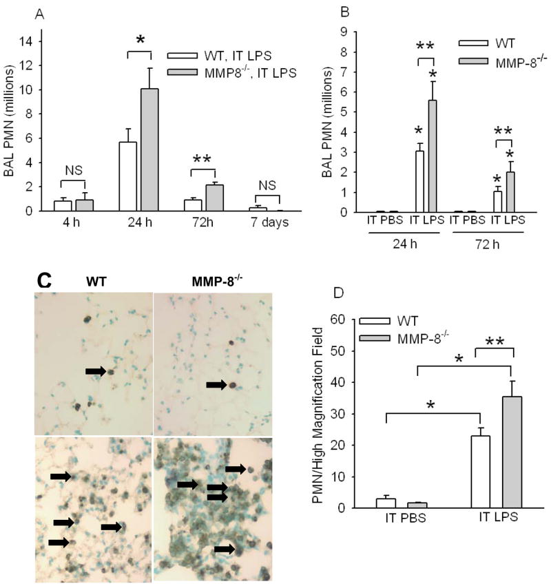
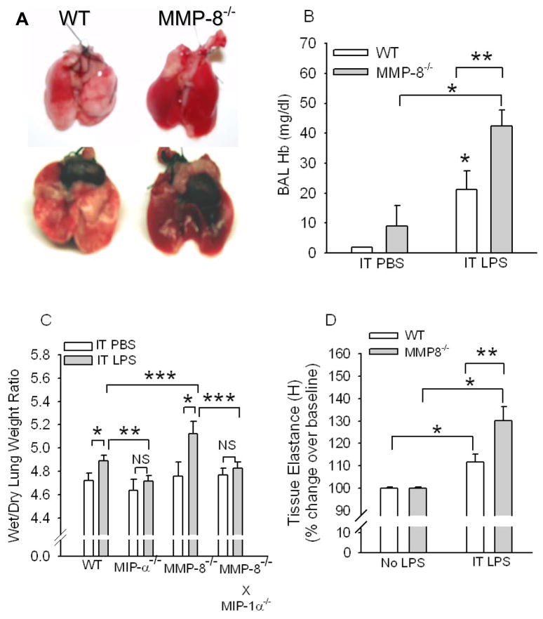
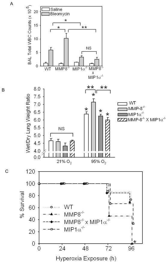
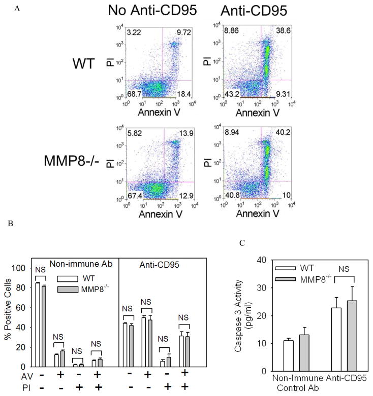
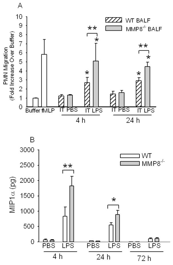
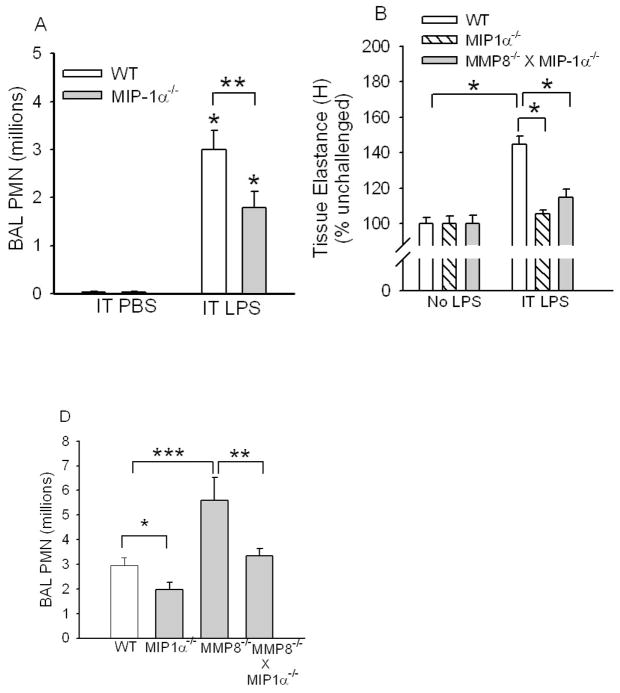
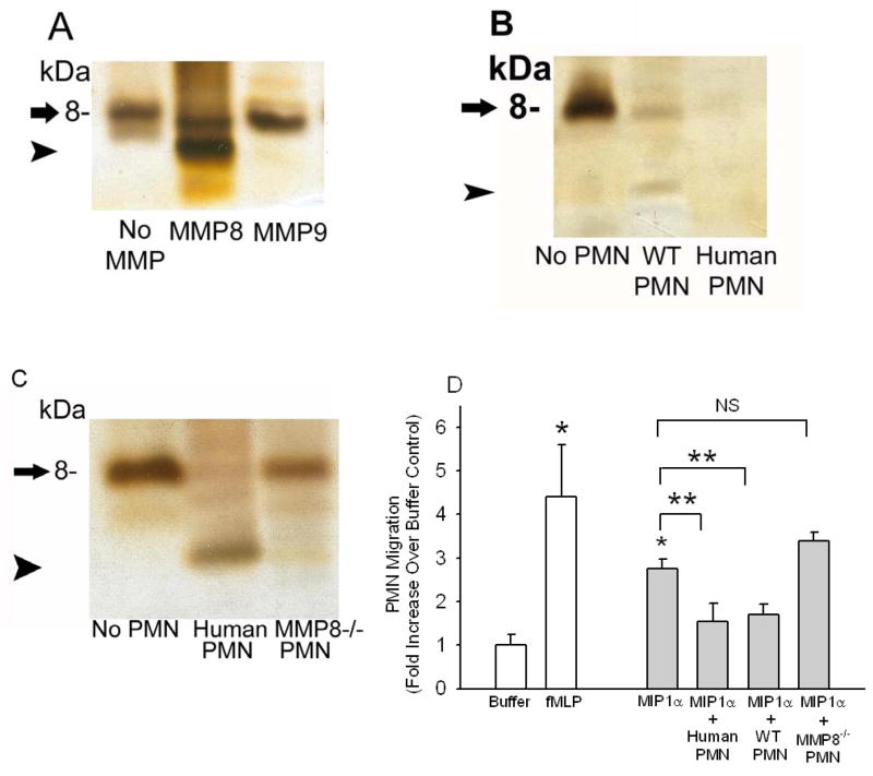
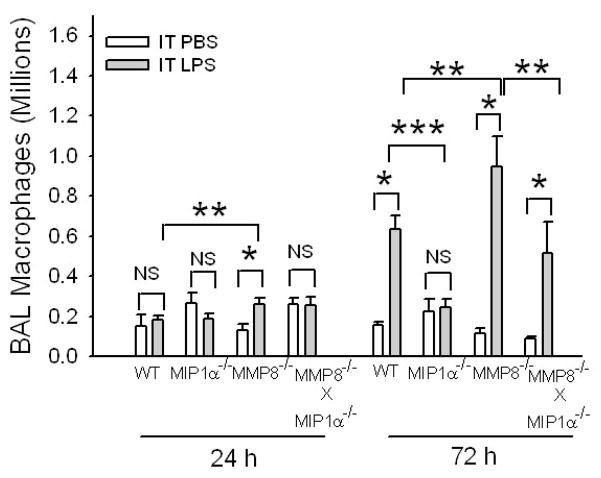
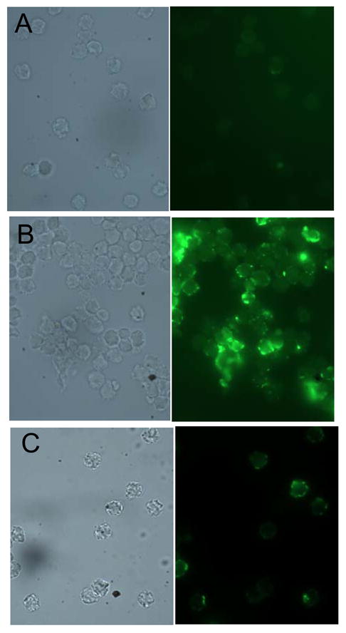
To determine the role of matrix metalloproteinase-8 (MMP-8) in acute lung injury (ALI), we delivered LPS or bleomycin by the intratracheal route to MMP-8(-/-) mice versus wild-type (WT) mice or subjected the mice to hyperoxia (95% O(2)) and measured lung inflammation and injury at intervals. MMP-8(-/-) mice with ALI had greater increases in lung polymorphonuclear neutrophils (PMNs) and macrophage counts, measures of alveolar capillary barrier injury, lung elastance, and mortality than WT mice with ALI. Bronchoalveolar lavage fluid (BALF) from LPS-treated MMP-8(-/-) mice had more MIP-1alpha than BALF from LPS-treated WT mice, but similar levels of other pro- and anti-inflammatory mediators. MIP-1alpha(-/-) mice with ALI had less acute lung inflammation and injury than WT mice with ALI, confirming that MIP-1alpha promotes acute lung inflammation and injury in mice. Genetically deleting MIP-1alpha in MMP-8(-/-) mice reduced the increased lung inflammation and injury and mortality in MMP-8(-/-) mice with ALI. Soluble MMP-8 cleaved and inactivated MIP-1alpha in vitro, but membrane-bound MMP-8 on activated PMNs had greater MIP-1alpha-degrading activity than soluble MMP-8. High levels of membrane-bound MMP-8 were detected on lung PMNs from LPS-treated WT mice, but soluble, active MMP-8 was not detected in BALF samples. Thus, MMP-8 has novel roles in restraining lung inflammation and in limiting alveolar capillary barrier injury during ALI in mice by inactivating MIP-1alpha. In addition, membrane-bound MMP-8 on activated lung PMNs is likely to be the key bioactive form of the enzyme that limits lung inflammation and alveolar capillary barrier injury during ALI.


Conflict of interest statement

Disclosures

None of the authors have conflicts of interest or financial interests to disclose.

Figures

Figure 1
Figure 1. MMP-8−/− mice have increased lung PMNs during LPS-mediated ALI
In A, WT and MMP-8−/− mice both in a mixed SvEv129 X C57BL/6J strain were given IT LPS (10 μg) or IT PBS. BAL was performed 4 h to 7 d later and PMN counts were determined. Data are mean ± SEM; n = 5–9 mice/group. Asterisk indicates p =0.015 and ** indicates p = 0.003. In B, to provide assurance that MMP-8 reduces acute lung inflammation in mice, PMN counts in BAL samples were quantified 24 h and 72 h after IT LPS (n = 8–14 mice/group) or IT PBS (4–8 mice/group) in MMP-8−/− mice backcrossed to the pure C57BL/6J background (F10 generation) or pure C57BL/6J WT littermate control mice. Data are mean ± SEM. Asterisk indicates p < 0.006 compared to mice of the same genotype given IT PBS at the same time point and ** indicates p ≤ 0.034. C shows representative lung sections from C57BL/6J WT littermate control mice (left panels) and C57BL/6J MMP-8−/− mice (right panels) 24 h after IT PBS (top panels) or IT LPS (bottom panels) immunostained for a PMN marker (Ly-6G). Arrows indicate Ly-6G-positive PMN. Magnification X 200. In D, Ly-6G-positive PMN were counted in eight consecutive 1000 X magnification fields in lung sections harvested from C57BL/6J WT littermate control mice or C57BL/6J MMP-8−/− mice 24 h after IT PBS (n = 4 mice/group) or IT LPS (n = 6–7 mice/group). Data are mean ± SEM. Asterisk indicates p ≤ 0.01 and ** indicates p = 0.047. No staining was observed in lung sections from WT or MMP-8−/− mice immunostained with an isotope-matched non-immune control antibody (not shown).
Figure 2
Figure 2. MMP-8−/− mice have greater LPS-induced lung injury than WT mice
In A, C57BL6J WT mice and C57BL/6J MMP-8−/− mice were given 10 μg of LPS by the IT route and 24 h later lungs were removed. Note the more extensive inflammation and hemorrhage in the lungs of the MMP-8−/− mouse (right) given IT LPS than in those from the LPS-treated WT mouse (left). Lungs from WT and MMP-8−/− mice given IT PBS had no obvious lung inflammation or hemorrhage (not shown). In B, hemoglobin levels were measured in BAL samples from WT and MMP-8−/− mice 24 h after IT PBS or LPS. Data are mean ± SEM; n = 3 for groups given IT PBS and 13 for groups given IT LPS. In C, wet/dry lung weight ratios were measured in WT C57BL/6J littermate control mice, C56BL/6J MIP-1α−/−, C57BL/6J MMP-8−/−, and C56BL/6J MMP-8−/− X MIP-1α−/− mice 24 h after IT LPS (n = 11–22 mice/group) or IT PBS (n = 5–9 mice/group). Data are mean ± SEM. Asterisk indicates p <0.001; ** indicates p < 0.03; and *** indicates p < 0.05. NS indicates not significantly different. In D, WT or MMP-8−/− mice (C57BL/6J strain) were given two doses of LPS (20 μg) by the IT route 24 h apart, and 24 h after the last dose of LPS, lung elastance (H which is a measure of lung stiffness) was measured in LPS-treated mice vs. unchallenged mice using a Flexivent apparatus. Data are mean ± SEM; n = 6–9 mice per group. Asterisk indicates p < 0.05 compared with unchallenged mice of the same genotype and ** indicates p = 0.017.
Figure 3
Figure 3. MMP-8−/− mice have greater lung inflammation and injury in other models of ALI
In A, WT, MMP-8−/−, MIP-1α−/−, and MMP-8−/− X MIP-1α−/− mice (all in the pure C57BL/6 strain) were given 75 mU bleomycin (n = 11–18 mice/group) or saline (n = 4–6 mice/group) by the IT route. After 7 days, WBC were counted in BAL samples. Data are mean ± SEM. Asterisk indicates p ≤ 0.027 and **, p < 0.001. In B, WT (n =15), MMP-8−/− (n = 13), MIP-1α−/− (n =9), and MMP-8−/− X MIP-1α−/− (n= 13) mice (all in the pure C57BL/6 strain) were exposed to 95% 02 for up to 96 h and survival was recorded. Asterisk indicates p < 0.001 compared to WT mice at 96 h. In C, the same genotypes described in B were exposed for 72 h to 95% 02 (n = 7–13 mice/group) or 21% 02 (n = 4–9 mice/group) and wet to dry lung weight ratios were measured. Data are mean ± SEM. Asterisk indicates p< 0.001 compared to the same genotype exposed to 21% O2 and ** indicates p ≤ 0.007.
Figure 4
Figure 4. MMP-8 does not regulate survival of PMNs induced to undergo apoptosis in vitro by serum starvation
In A-C, PMNs were isolated from the bone marrow of WT and MMP-8−/− mice and aliquots of the cells were induced to undergo apoptosis by incubating the cells in the absence of serum or agonists to trigger the intrinsic apoptosis pathway. Freshly isolated cells (no serum starvation) or cells serum starved for 18 h were stained with annexin-V conjugated to Alexa 488 or propidium iodide (A and B) or active caspase 3 was measured in cell lysates as described in Methods (C). Representative FACS plots are shown in A and quantitative data for annexin V and propodium iodide staining in B. In B and C, data are mean ± SEM (n = 4 mice/group).
Figure 5
Figure 5. MMP-8 does not regulate survival of PMNs induced to undergo apoptosis in vitro by FAS (CD95) activation
In A-C, PMNs were isolated from the bone marrow of WT and MMP-8−/− mice and aliquots of the cells were induced to undergo apoptosis by incubating the cells for 4 h at 37°C with equal amounts of anti-FAS (anti-CD95) to ligate and activate FAS death domain-containing receptors on PMNs. Cells were then stained with annexin-V conjugated to Alexa 488 or propidium iodide (A and B) or active caspase 3 was measured in cell lysates as described in Methods (C). Representative FACS plots are shown in A and quantitative data for annexin V and propodium iodide staining in B. In B and C, data are mean ± SEM (n = 4 mice/group).
Figure 6
Figure 6
MMP-8 reduces lung PMN chemokine activity and lung MIP-1α levels during LPS-mediated ALI in mice. In A, cell-free BALF samples from WT or MMP-8−/− mice 4 h or 24 h after IT PBS or IT LPS were tested for PMN chemotactic activity in triplicate using Boyden microchemotaxis assay chambers, along with buffer and 10−7M fMLP as controls (white bars). PMNs migrating through the membranes were counted in 6–10 consecutive 1000 X magnification fields for each well. Data are mean ± SEM. Asterisk indicates p ≤ 0.036 compared with BALF from the same genotype of mice given IT PBS at the same time point and ** indicates p ≤ 0.035. B. MIP-1α levels were measured in BALF samples from WT and MMP-8−/− mice 4–72 h after IT PBS (n = 5–11 mice/group) or IT LPS (9–23 mice/group) by ELISA. Data are mean ± SEM. Asterisk indicates p < 0.04 and ** indicates p = 0.032.
Figure 7
Figure 7. MIP-1α promotes LPS-mediated acute lung inflammation and injury and neutralization or genetic deletion of MIP-1α in MMP-8−/− mice reduces lung PMN counts in LPS-treated MMP-8−/− mice
In A, C57BL6J WT mice and C57BL/6J MIP-1α−/− mice were given PBS (n = 4 mice/group) or 10 μg LPS (n = 12–15 mice/group) by the IT route and 24 h later PMN were counted in BAL samples. Data are mean ± SEM. Asterisk indicates p < 0.003 compared to mice of the same genotype given IT PBS and ** indicates p = 0.038. In B, WT, MIP-1α−/−, and MMP-8−/− X MIP-1α−/− mice were not treated or given two doses of LPS (20 μg) by the IT route 24 h apart and 24 h after the last dose of LPS lung elastance (H) was measured using a Flexivent apparatus. Data are mean ± SEM; n = 7–17 mice/group. Asterisk indicates p < 0.001. There was no statistically significant difference in H between unchallenged MIP-1α−/− mice and LPS-treated MIP-1α−/− mice, or between unchallenged MMP-8−/− X MIP-1α−/− mice and LPS-treated MMP-8−/− X MIP-1α−/− mice, or between LPS-treated MMP-8−/− X MIP-1α−/− mice and LPS-treated MIP-1α−/− mice. In C, WT, MIP-1α−/−, MMP-8−/−, and MMP-8−/− X MIP-1α−/− mice were given 10 μg of LPS or PBS by the IT route, and 24 h later PMNs were counted in BAL samples. Data are mean ± SEM. Asterisk indicates p = 0.047; ** indicates p = 0.04; and *** indicates p = 0.002; n = 12–26 mice/group. PMNs were not detected in BAL samples from any mice given IT PBS (not shown).
Figure 8
Figure 8. MMP-8 cleaves and inactivates MIP-1α in vitro
Equal amounts of recombinant MIP-1α (2.5 μM in each lane) were incubated at 37°C for 18 h with and without 500 nM MMP-8 or 500 nM MMP-9 in A, or in B and C with or without (lane 1; No PMN) or with equal numbers (5 × 10⁶ cells/assay) of activated and fixed human PMNs (human PMN), activated and fixed WT murine PMNs (WT PMN), or activated and fixed MMP-8−/− murine PMNs (MMP-8−/− PMN). In A-C, reduced cell-free reaction products were analyzed on silver-stained 16.5% Tris-tricine gels. Arrow indicates intact MIP-1α and arrowhead indicates a cleavage product of MIP-1α. In D, rMIP-1α (2.5 μM) was incubated at 37°C for 18 h with and without fixed, activated human PMNs, WT PMNs or MMP-8−/− PMNs as described above. Residual MIP-1α-mediated PMN chemotactic activity was tested in cell-free supernatants samples in triplicate using Boyden microchemotaxis assay chambers (along with buffer alone or 10−7M fMLP as assay controls). Data are mean ± SEM; n = 3–5 separate samples/group. Asterisk indicates p = 0.029 compared to buffer alone and ** indicates p < 0.037.
Figure 9
Figure 9. MMP-8 reduces macrophage accumulation during LPS-mediated ALI
WT, MIP-1α−/−, MMP-8−/−, and MMP-8−/− X MIP-1α−/− mice were given 10 μg of LPS (n = 7–25 mice) or PBS (n = 5–10 mice) by the IT route and after 24–72 h, macrophages were counted in BAL samples. Data are mean ± SEM. Asterisk indicates p < 0.016; ** indicates p ≤ 0.05; and *** indicates p = 0.012.
Figure 10
Figure 10. Lung PMNs from WT mice given IT LPS express membrane-bound MMP-8
A WT mouse (A and B) and an MMP-8−/− mouse (C) were given 10 μg of LPS by the IT route, and 24 h later PMNs were isolated from their lungs by BAL. Cells were fixed and non-permeabilized cells immunostained with a rabbit anti-MMP-8 IgG (B and C) or a non-immune rabbit IgG (A) followed by goat anti-rabbit IgG conjugated to Alexa-488. Note the intense staining for MMP-8 on the surface of WT BAL PMNs stained with anti-MMP-8 (B) but minimal staining associated with WT PMNs stained with the control antibody (A) or with MMP-8−/− PMNs incubated with the anti-MMP-8 antibody (C).

Similar articles

Cited by

References

    1. Owen CA, Campbell EJ. The cell biology of leukocyte-mediated proteolysis. J Leukoc Biol. 1999;65:137–150. - PubMed
    1. Owen CA, Hu Z, Lopez-Otin C, Shapiro SD. Membrane-bound matrix metalloproteinase-8 on activated polymorphonuclear cells is a potent, tissue inhibitor of metalloproteinase-resistant collagenase and serpinase. J Immunol. 2004;172:7791–7803. - PubMed
    1. Balbin M, Fueyo A, Knauper V, Pendas AM, Lopez JM, Jimenez MG, Murphy G, Lopez-Otin C. Collagenase 2 (MMP-8) expression in murine tissue-remodeling processes. Analysis of its potential role in postpartum involution of the uterus. J Biol Chem. 1998;273:23959–23968. - PubMed
    1. Balbin M, Fueyo A, Tester AM, Pendas AM, Pitiot AS, Astudillo A, Overall CM, Shapiro SD, Lopez-Otin C. Loss of collagenase-2 confers increased skin tumor susceptibility to male mice. Nat Genet. 2003;35:252–257. - PubMed
    1. Cowland JB, Borregaard N. The individual regulation of granule protein mRNA levels during neutrophil maturation explains the heterogeneity of neutrophil granules. J Leukoc Biol. 1999;66:989–995. - PubMed

Publication types

MeSH terms