Comparative Study
. 2010 Feb 1;184(3):1168-79.
doi: 10.4049/jimmunol.0902750. Epub 2009 Dec 30.

Cell type-specific recognition of human metapneumoviruses (HMPVs) by retinoic acid-inducible gene I (RIG-I) and TLR7 and viral interference of RIG-I ligand recognition by HMPV-B1 phosphoprotein

Affiliations
Comparative Study

Cell type-specific recognition of human metapneumoviruses (HMPVs) by retinoic acid-inducible gene I (RIG-I) and TLR7 and viral interference of RIG-I ligand recognition by HMPV-B1 phosphoprotein

Nadege Goutagny et al. J Immunol. .

Abstract

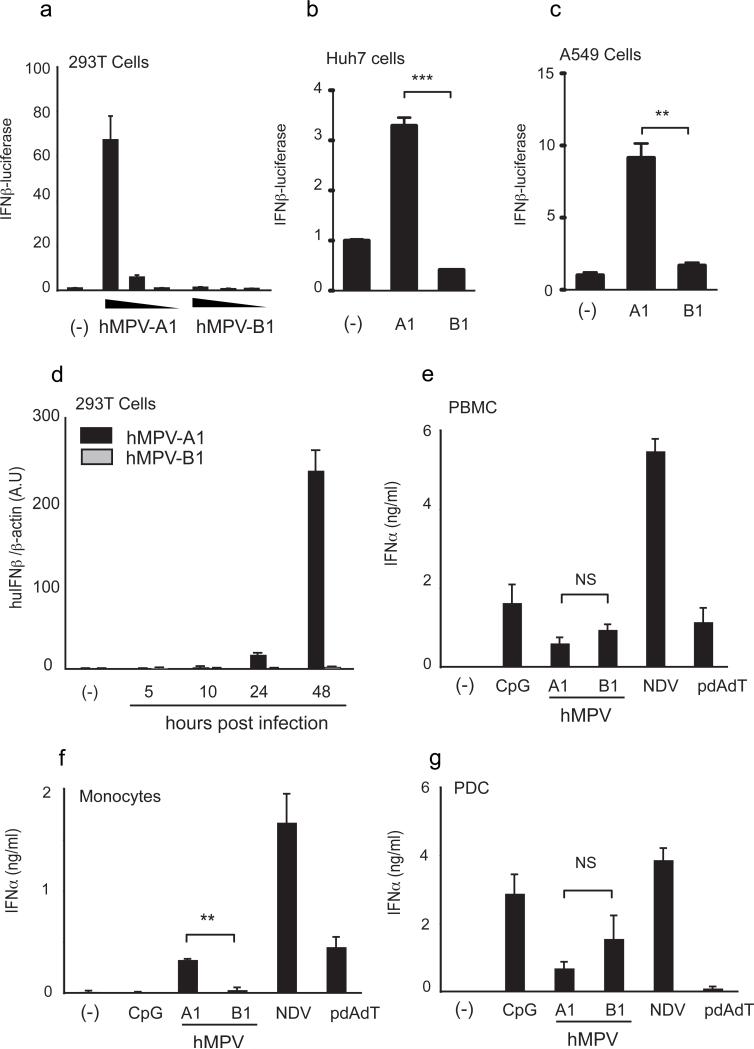
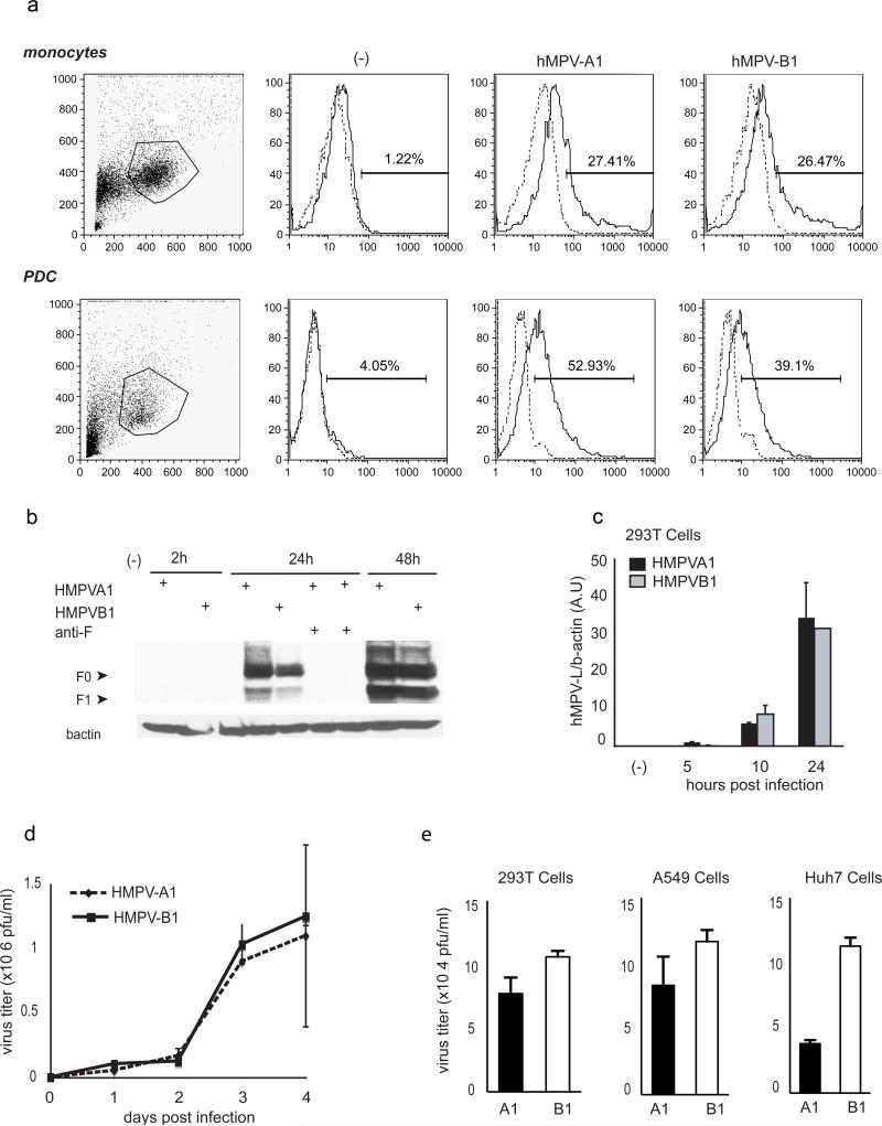
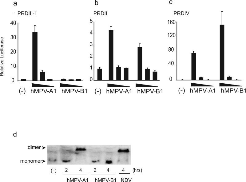
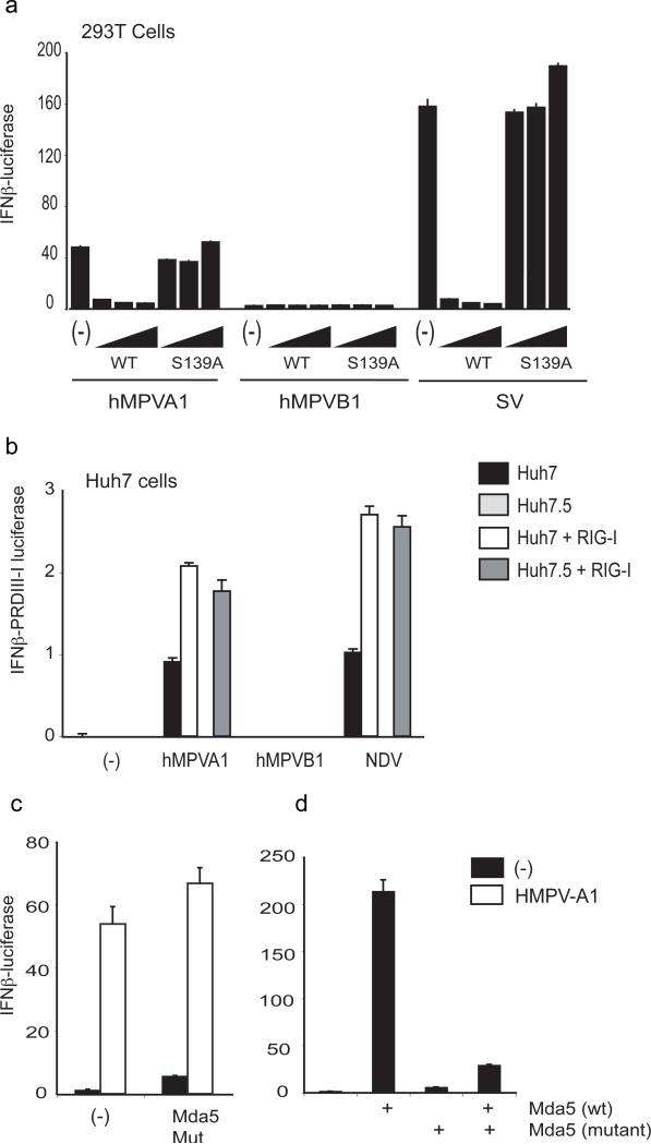
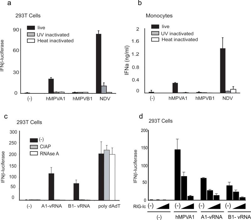
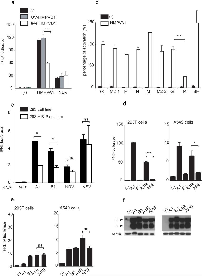
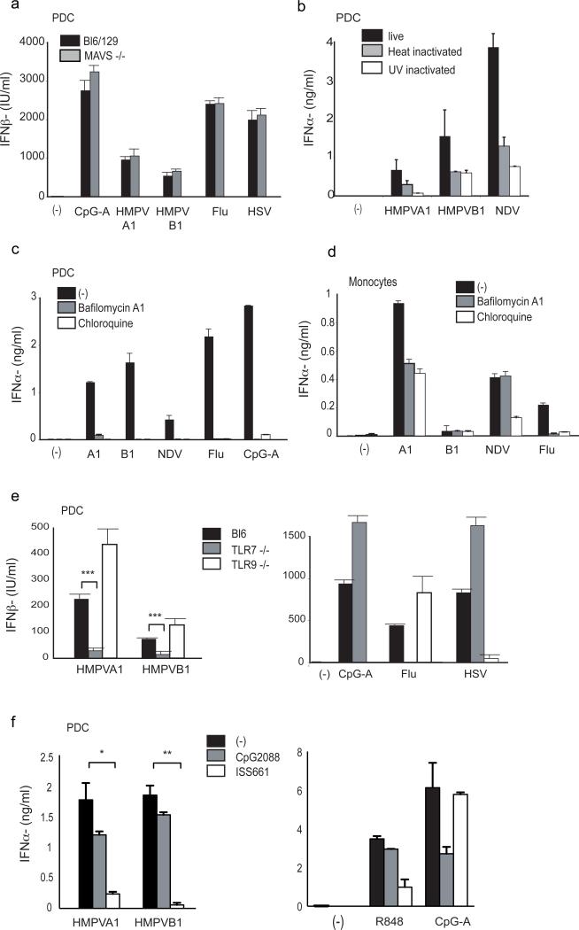
Human metapneumoviruses (HMPVs) are recently identified Paramyxoviridae that contribute to respiratory tract infections in children. No effective treatments or vaccines are available. Successful defense against virus infection relies on early detection by germ line-encoded pattern recognition receptors and activation of cytokine and type I IFN genes. Recently, the RNA helicase retinoic acid-inducible gene I (RIG-I) has been shown to sense HMPV. In this study, we investigated the abilities of two prototype strains of HMPV (A1 [NL\1\00] and B1 [NL\1\99]) to activate RIG-I and induce type I IFNs. Despite the abilities of both HMPV-A1 and HMPV-B1 to infect and replicate in cell lines and primary cells, only the HMPV-A1 strain triggered RIG-I to induce IFNA/B gene transcription. The failure of the HMPV-B1 strain to elicit type I IFN production was dependent on the B1 phosphoprotein, which specifically prevented RIG-I-mediated sensing of HMPV viral 5' triphosphate RNA. In contrast to most cell types, plasmacytoid dendritic cells displayed a unique ability to sense both HMPV-A1 and HMPV-B1 and in this case sensing was via TLR7 rather than RIG-I. Collectively, these data reveal differential mechanisms of sensing for two closely related viruses, which operate in cell type-specific manners.


Figures

Figure 1
Figure 1. HMPV A1 and B1 strains differentially induce IFNβ gene transcription
(a-c) 293T, Huh7 and A549 cells were transfected with the full length IFNβ promoter. Cells were infected with HMPV-A1 or HMPV-B1 (from 5×10⁴ to 5×10²pfu/ml for 293T, 5×10⁴pfu/ml for other lines) for an additional 24h. Data are expressed as fold induction relative to the reporter-only control and are the mean ± SD. (d) 293T cells were stimulated with HMPV-A1 or HMPV-B1 (5.104pfu/ml) for the indicated time. Levels of human IFNβ and β-actin were quantified in RNA samples by real-time PCR. Results are presented in arbitrary units as the ratio of IFNβ over 100 copies of β-actin. (e-g) Total PBMC, monocytes or PDC were stimulated with HMPV-A1, HMPV-B1 (2×10⁵pfu/ml), CpG-A 2216 (3μM), NDV (8HAU/ml), or poly(dAdT)•poly(dAdT) (5μg/ml) for 24h. Protein levels were measured in the supernatant of culture by ELISA and presented as the mean ± SD.
Figure 2
Figure 2. HMPV-A1 and HMPV-B1 infect and replicate in primary cells and cell lines
(a) Purified monocytes (6×10⁵ cells/well) and PDC (2.105 cells/well) were cultured for 24h in presence of HMPV-A1 and B1 (3.2×10⁵pfu/ml). Cells were harvested and stained for specific cell surface antigen (CD14 or BDCA2) and F protein expression using an anti-F biotinylated antibody and PE or APC-conjugated steptavidin. Results are presented as histogram overlays of anti-F antibody staining versus isotype control. Displayed percentages were substracted from the isotype control. (b). Infection of 293T cells by HMPV was assessed by immunoblot analysis. Cells (6×10⁵cells/well) were infected with HMPV-A1 or HMPV-B1 (5×10⁴pfu/ml) for 2 to 48h. A specific anti-F biotinylated antibody and HRP-conjugated steptavidin were used to detect F protein in cell lysates. F0 and F1 forms of the F protein are indicated by an arrow. Anti-F #338 neutralizing antibody was used to show the specificity. The lower panel shows β-actin levels in the samples. (c) Replication of HMPV in 293T was quantified by real-time PCR. Cells were infected with HMPV-A1 or HMPV-B1 (5×10⁴pfu/ml) for the indicated time points. Levels of HMPV-L gene and human β-actin were quantified in RNA samples using specific primers. Results are presented in arbitrary unit as the ratio of HMPV-L gene over 100 copies of β-actin. (d-e) Virus titers were determined after infection of several cell types (3×10⁵/well) with HMVPA1 or B1 strain (105pfu/ml). Virus titers were measured for the indicated time points after infection in Vero cells (d) or after 24h post infection in 293T, A549 and Huh7 cells (e) and presented as pfu/ml.
Figure 3
Figure 3. Differential activation of IRF3 by the HMPV A1 and B1 strains
(a-c) 293T cells were transfected with the IFNB PRDIII-I, PRDII or PRDIV reporter genes as detailed in Material and Methods. Cells were infected with HMPV-A1 or HMPV-B1 (range from 5×10⁴ to 5×10²pfu/ml) for an additional 24h. Data are expressed as fold induction relative to the reporter-only control and are the mean ± SD. (d) A549 cells were stimulated with viruses for the indicated period of time. Nuclear protein were extracted and analyzed by native PAGE. The monomer and dimeric forms of IRF3 are indicated by arrows.
Figure 4
Figure 4. HMPV-A1 induces IFNβ via the RIG-I/MAVS pathway
(a) 293T cells were transfected with the IFNβ reporter gene and increasing concentrations (5-80ng/well) of the WT or S139A inactive NS3/4A protease from HCV. Cells were stimulated for an additional 24h with HMPV-A1, HMPV-B1 (5×10⁴pfu/ml) or SV (400HAU/ml). Data are expressed as fold induction relative to the reporter-only control and are the mean ± SD. (b) Parental human hepatoma cell line Huh7 and Huh7.5 were transfected with the IFNβ reporter gene in the absence or presence of pEFBos-huRIG-I Flag (40ng/well). Cells were stimulated with HMPV-A1, HMPV-B1 (5×10⁴pfu/ml) or NDV (64HAU/ml) for an additional 24h. Results are normalized by renilla luciferase and presented as arbitrary units after correction between the 2 cell lines using the pGL3-control. (c) 293T cells were transfected with the IFNβ reporter gene and (d) WT or a dominant negative mutant of MDA-5 (80 ng/well). Cells were stimulated with HMPV-A1 (5×10⁴pfu/ml) for additional 24hrs. Data are expressed as fold induction relative to the reporter-only control and are the mean ± SD.
Figure 5
Figure 5. Induction of IFNβ by HMPV-A1 requires viral replication
(a) 293T cells were transfected with the full length IFNβ promoter. Cells were stimulated 24h later with live, UV- or heat-inactivated HMPV-A1, HMPV-B1 (2×10⁵pfu/ml) or NDV (8HAU/ml) for an additional 24h. Data are expressed as fold induction relative to the reporter-only control and are the mean ± SD. (b) Freshly isolated monocytes were stimulated for 24h with live, UV- or heat-inactivated HMPV-A1, HMPV-B1 (2×10⁵pfu/ml) or NDV (8HAU/ml). Human IFNα protein levels were measured in the supernatant of culture by ELISA and presented as the mean ± SD. (c) 293T cells were transfected with viral RNA purified from HMPV-A1 or B1 (80ng), or poly(dAdT)•poly(dAdT) (20ng) along with the IFNβ and TK-renilla reporter genes. vRNAs and poly(dAdT)•poly(dAdT) were treated with calf intestinal alkaline phosphatase (CIAP) or RNAse A prior to stimulation as indicated. (d) 293T cells were transfected with increasing amounts of RIG-IC (0-2-20ng/well) along with the IFNB reporter and vRNAs (80ng), or stimulate with HMPV-A1 (5×10⁴pfu/ml). In all cases, luciferase activity was measured 24h post-transfection and data are expressed as fold induction relative to the reporter-only control and are the mean ± SD.
Figure 6
Figure 6. HMPV-B1 P protein prevents RIG-I from sensing HMPV vRNA
(a) 293T cells were transfected with the IFNβ reporter gene one day prior to stimulation. Cells were pre-incubated with live or UV (2J/cm2) inactivated HMPV-B1 (105pfu/ml) for 24 hours. Cells were then stimulated with HMPV-A1 (5×10⁴pfu/ml) or NDV (8HAU) for an additional 24hours. Results are normalized by renilla luciferase and presented as fold induction relative to the reporter-only control and are the mean ± SD. (b). 293T cells were transfected with the IFNβ PRDIII-I reporter gene and 40ng of plasmids encoding HMVB1 proteins as indicated. Cells were stimulated for an additional 24h with HMPV-A1 (5×10⁴pfu/ml). Data were normalized by renilla luciferase and are expressed as a percentage of activation relative to conditions with the virus in the absence of exogenous proteins and are the mean ± SD. (c) HEK293 cells and HEK293 cells stably expressing the P protein from HMPV-B1 were transfected with the full length IFNβ reporter gene along with the viral RNA (80ng) purified from HMPV-A1, B1, NDV, or VSV, or with total RNA (80ng) purified from Vero cells as control. Results are normalized by renilla and pGL3 control luciferase, and presented as mean ± SD. (d-f). 293T and A549 cells were transfected with the full length IFNB or the PRDIV reporter along with the TK-renilla reporter gene, one day prior to stimulation. Cells were infected for 24 hours with the following viruses: HMPV-A1, HMPV-B1, HMPV-A1 recovered from cDNA (A1R), or the chimeric HMPV-A1 virus encoding the P protein from the B1 strain (APB) (5×10⁴pfu/ml). Results are normalized by renilla luciferase and are presented as fold induction relative to the reporter-only control and are the mean ± SD. A western blot of F protein levels is shown (f).
Figure 7
Figure 7. Induction of type I IFN in PDC in response to both HMPV-A1 and B1 is TLR7-mediated
(a). PDC from C57Bl6/129 and MAVS−/− mice were stimulated for 24h with CpG-A (2μM), Heat-inactivated Influenza (MOI=0.1), HSV (MOI=100) HMPV-A1, HMPV-B1 (2×10⁵pfu/ml). Mouse IFNβ protein levels were measured in the supernatant of culture by ELISA and presented as the mean ± SD. (b). Freshly isolated PDC were stimulated for 24h with live, UV- or heat-inactivated HMPV-A1, HMPV-B1 (2×10⁵pfu/ml) or NDV (8HAU/ml). Human IFNα protein levels were measured in the supernatant by ELISA and presented as the mean ± SD. (c-d) Freshly isolated PDC and monocytes were stimulated with HMPV-A1, HMPV-B1 (2×10⁵pfu/ml), NDV (8HAU/ml), Heat-inactivated Influenza (MOI=0.1), CpG-A (2μM) for 24 hours. Cells were pre-incubated with chloroquine (1μM) or bafilomycin A1 (4μM for PDC, 10μM for monocytes) for 1 hour were indicated. Human IFNα protein levels were measured in the supernatant by ELISA and presented as the mean ± SD. (e). Mouse PDC from C57Bl6, TLR7−/− and TLR9−/−mice were stimulated for 24h with CpG-A (2μM), Heat-inactivated Influenza (MOI=0.1), HSV (MOI=100) HMPV-A1, HMPV-B1 (2×10⁵pfu/ml). Mouse IFNβ protein levels were measured by ELISA and presented as the mean ± SD. (f) Freshly isolated PDC were stimulated with HMPV-A1, HMPV-B1 (2×10⁵pfu/ml), R848 (10μM), CpG-A (2μM) for 24 hours. Cells were pre-incubated with CpG2088 (0.1μM) or ISS661 (2μM) for 1 hour were indicated. Human IFNα protein levels were measured in the supernatant by ELISA and presented as the mean ± SD.

Similar articles

Cited by

References

    1. van den Hoogen BG, de Jong JC, Groen J, Kuiken T, de Groot R, Fouchier RA, Osterhaus AD. A newly discovered human pneumovirus isolated from young children with respiratory tract disease. Nat Med. 2001;7:719–724. - PMC - PubMed
    1. van den Hoogen BG, Herfst S, Sprong L, Cane PA, Forleo-Neto E, de Swart RL, Osterhaus AD, Fouchier RA. Antigenic and genetic variability of human metapneumoviruses. Emerg Infect Dis. 2004;10:658–666. - PMC - PubMed
    1. Ji W, Chen ZR, Wang YQ. [Comparison of the clinical manifestation and lung function between RSV and hMPV lower respiratory tract infection]. Zhonghua Er Ke Za Zhi. 2009;47:71–73. - PubMed
    1. van den Hoogen BG, Bestebroer TM, Osterhaus AD, Fouchier RA. Analysis of the genomic sequence of a human metapneumovirus. Virology. 2002;295:119–132. - PubMed
    1. Huck B, Scharf G, Neumann-Haefelin D, Puppe W, Weigl J, Falcone V. Novel human metapneumovirus sublineage. Emerg Infect Dis. 2006;12:147–150. - PMC - PubMed

Publication types

MeSH terms