. 2010 Mar 5;285(10):7776-83.
doi: 10.1074/jbc.M109.077156. Epub 2009 Dec 30.

Differentiation of embryonic stem cells 1 (Dies1) is a component of bone morphogenetic protein 4 (BMP4) signaling pathway required for proper differentiation of mouse embryonic stem cells

Affiliations

Differentiation of embryonic stem cells 1 (Dies1) is a component of bone morphogenetic protein 4 (BMP4) signaling pathway required for proper differentiation of mouse embryonic stem cells

Luigi Aloia et al. J Biol Chem. .

Abstract

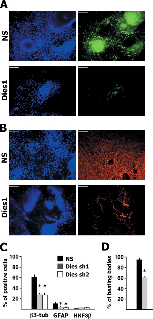
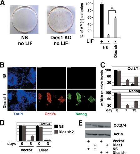
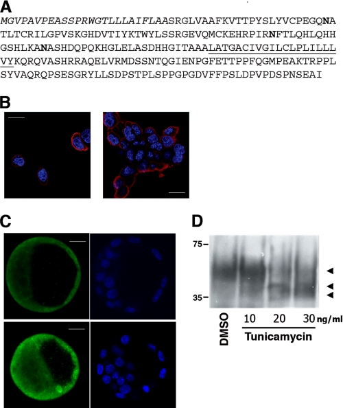
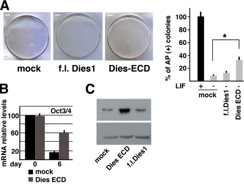
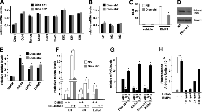
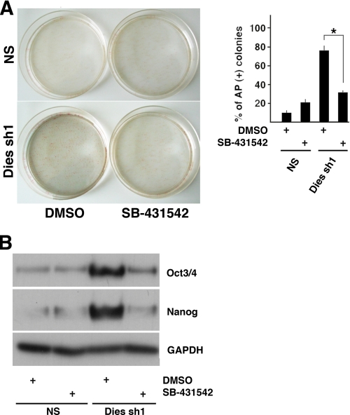
Embryonic stem cells (ESCs) are pluripotent cells able to grow indefinitely in culture and to differentiate into all cell types of embryos upon specific stimuli. Molecular mechanisms controlling the unique characteristics of ESCs are still largely unknown. We identified Dies1 (Differentiation of ESCs 1), an unpublished gene, that encodes a type I membrane protein. ESCs stably transfected with Dies1 small hairpin RNAs failed to properly differentiate toward neural and cardiac cell fate upon appropriate stimuli and continued to express markers of undifferentiated cells, such as the membrane-associated alkaline phosphatase, and transcription factors, like Oct3/4 and Nanog, when grown under conditions promoting differentiation. Our results demonstrated that Dies1 is required for BMP4/Smad1 signaling cascade; in undifferentiated ESCs Dies1 knockdown did not affect the expression of leukemia inhibitory factor downstream targets, whereas it resulted in a strong decrease of BMP4 signaling, as demonstrated by the decrease of Id1, -2, and -3 mRNAs, the decreased activity of Id1 gene promoter, and the reduced phospho-Smad1 levels. Dies1 knockdown had no effect in murine ESCs when the expression of the BMP4 receptor Alk3 was suppressed. The phenotype induced by Dies1 suppression in ESCs is due to the indirect activation of the Nodal/Activin pathway, which is a consequence of the BMP4 pathway inhibition and is sufficient to support the mESC undifferentiated state in the absence of leukemia inhibitory factor.


Figures

FIGURE 1.
FIGURE 1.
Dies1 silencing impairs mouse ESC differentiation. A, E14 mouse ESCs stably transfected with a construct driving the expression of EGFP under the control of the neuron-specific α1-tubulin gene promoter were transfected with an shRNA targeting the Dies1 gene. 13 days after the induction of differentiation, ESCs transfected with the nonsilencing (NS) shRNA showed many cells expressing EGFP (upper right panel), although Dies1 suppressed cells did not (lower right panel). Scale bars, 100 μm. B, E14Tg2A ESCs were transfected with the shRNA targeting Dies1. 13 days after the induction of differentiation, ESCs transfected with the control NS shRNA showed most of the cells expressing β3-tubulin (upper right panel), whereas only a few Dies1 KD cells were decorated by the β3-tubulin antibody. C, mouse ESCs were transfected with NS shRNA (black bars) or with two different shRNAs targeting Dies1 (Dies sh1, gray bars, and Dies sh2, white bars). 13 days after the induction of the differentiation, cells were stained with various antibodies, and positive cells were counted. The histogram reports the percentages of cells positive for antibodies recognizing the neuron-specific β3-tubulin (β3-tub), glial fibrillary acidic protein (GFAP), and hepatocyte nuclear factor 3β (HNF3β). Values are the means of three independent experiments plus S.D. *, p ≤ 0.001. D, mouse ESCs transfected with NS (black bar) or with Dies sh1 (gray bar) were cultured as embryoid bodies in a selected serum for 8 days to obtain differentiation into cardiomyocytes. The percentage of embryoid bodies containing spontaneously contracting clusters (beating hearts) was measured in three independent experiments. S.D. is indicated. *, p ≤ 0.001.
FIGURE 2.
FIGURE 2.
Dies1 suppression maintains ESCs in undifferentiated state in the absence of LIF. A, ESCs stably transfected with NS or Dies1 silencing shRNAs were grown for 6 days in the absence of LIF and then stained for AP. This marker of ESC undifferentiated state was clearly observed in more than 50% of the colonies of Dies1 KD cells, although only about 10% of the NS shRNA transfected colonies were AP-positive. The experiments were done in triplicate. *, p ≤ 0.001. B, Dies1 KD ESCs grown 13 days in neural differentiation medium still expressed Oct3/4 and Nanog, although NS-transfected cells are completely negative for these master genes of stemness. Scale bars, 250 μm. C, mRNA levels of Oct3/4 and Nanog were measured in cells cultured as in A. The two mRNAs were strongly reduced in NS shRNA-transfected cells (black bars), although they were clearly detectable at 7 and 13 days after the induction of differentiation in Dies1 KD cells (gray bars). D, expression of a Dies1 cDNA resistant to the shRNA used to silence endogenous Dies1 rescued the phenotype of Dies1 KD ESCs. In cells expressing recombinant Dies1 (gray bars), Oct3/4 mRNA was expressed at the same levels of controls (black bars). E, as in D, Oct3/4 protein levels are rescued by Dies1 overexpression.
FIGURE 3.
FIGURE 3.
Dies1 is a membrane protein. A, Dies1 amino acid sequence predicted from mouse Dies1 gene. N-terminal signal peptide is in italic. Three possible Asn glycosylation sites are in boldface. Putative transmembrane tract is underlined. B, confocal images demonstrating that Dies1-FLAG is localized at the plasma membrane of ESCs, in isolated cells (left panels), and in clusters (right panel). Scale bars, 50 μm. C, confocal images of mouse blastocysts stained with Dies1 antibody, which indicate that Dies1 is mainly located on the cell surface. Scale bars, 20 μm. D, Western blot analysis of Dies1-FLAG from ESCs treated with the indicated different concentrations of tunicamycin to inhibit N-glycosylation. The control lane refers to cells treated with DMSO.
FIGURE 4.
FIGURE 4.
Dominant negative effect of Dies1 extracellular domain. A, ESCs stably transfected with full-length (f.l.) Dies1 or with Dies1 extracellular domain (Dies-ECD) were grown for 6 days in the absence of LIF and then stained for AP. 11% of colonies expressing full-length Dies1 are AP-positive, versus 33% of the Dies-ECD-expressing colonies. The experiments were done in triplicate. *, p ≤ 0.01. B, mRNA levels of Oct3/4 were measured in cells transfected as in A with Dies-ECD. Oct3/4 mRNA is clearly detectable at 6 days after LIF withdrawal. C, Oct3/4 protein in cells transfected with full-length (f.l.) Dies1, Dies-ECD, or empty vector (mock) after 6 days without LIF.
FIGURE 5.
FIGURE 5.
Dies1 suppression inhibits BMP4 signaling pathway. A, mRNA levels of the indicated genes were measured on total RNA by real time PCR. Dies1 KD was obtained with two different shRNAs (Dies sh1 and Dies sh2). B, mRNA levels of genes downstream of the BMP4 signaling pathway. All the Id mRNAs are significantly reduced in Dies1 KD cells compared with nonsilenced cells. All values are significantly (p < 0.01) different from the mean values in NS-transfected cells. C, ESCs stably transfected with NS or Dies1 silencing shRNAs were treated or not with BMP4 and transiently transfected with a vector where the luciferase gene was under the control of the Id1 gene promoter. The values are expressed as fold changes of luciferase/Renilla activity. D, Western blot analysis of ESCs transfected with NS or Dies1 silencing shRNA. The antibodies used recognize Smad1 and the phosphorylated forms of Smads 1/5/8. E, mRNA levels of genes downstream of the Nodal/Activin signaling pathway. F, ESCs stably transfected with NS or Dies1 silencing shRNAs were treated with SB-431542 that inhibits the Nodal/Activin signaling and with DMSO as a control. Id1, Id3, and Lefty1 mRNAs were measured by real time PCR. G, ESCs were transiently transfected with Dies1 shRNAs or Alk3 small interfering RNA or both. Alk3, Id3, and Lefty1 mRNAs were measured by real time PCR. H, BMP4 binds to Dies1 extracellular domain. Europium-labeled BMP4 was incubated with Strep-tagged Dies1 extracellular domain. Unlabeled BMP4 was mixed with the labeled compound at the indicated concentrations. Gray bar represents the fluorescence value measured when labeled BMP4 was incubated in wells not coated with Dies1 ECD (incubated with control conditioned medium of HEK293). Black bars refer to fluorescence of wells coated with Strep-tagged Dies1 ECD. All the experiments were done at least in triplicate. *, p ≤ 0.01; **, p ≤ 0.001.
FIGURE 6.
FIGURE 6.
Inhibition of Nodal/Activin signaling pathway masks the effects of Dies1 silencing in ESCs. A, ESCs stably transfected with NS or Dies1 silencing shRNAs were grown for 6 days in the absence of LIF and then stained for AP. Cells were treated with SB-431542 or with DMSO as a control. % of colonies AP-positive was calculated in triplicate experiments. *, p ≤ 0.001. B, Western blot analysis of Oct3/4 and Nanog in the cells treated as described in A.

Similar articles

Cited by

References

    1. Boiani M., Schöler H. R. (2005) Nat. Rev. Mol. Cell Biol. 6, 872–884 - PubMed
    1. Niwa H. (2007) Development 134, 635–646 - PubMed
    1. Loh Y. H., Wu Q., Chew J. L., Vega V. B., Zhang W., Chen X., Bourque G., George J., Leong B., Liu J., Wong K. Y., Sung K. W., Lee C. W., Zhao X. D., Chiu K. P., Lipovich L., Kuznetsov V. A., Robson P., Stanton L. W., Wei C. L., Ruan Y., Lim B., Ng H. H. (2006) Nat. Genet. 38, 431–440 - PubMed
    1. Chambers I., Tomlinson S. R. (2009) Development 136, 2311–2322 - PMC - PubMed
    1. Smith A. G., Heath J. K., Donaldson D. D., Wong G. G., Moreau J., Stahl M., Rogers D. (1988) Nature 336, 688–690 - PubMed

Publication types

MeSH terms