. 2010 Feb 26;285(9):6848-56.
doi: 10.1074/jbc.M109.074807. Epub 2009 Dec 30.

Filling the gap, evolutionarily conserved Omp85 in plastids of chromalveolates

Affiliations

Filling the gap, evolutionarily conserved Omp85 in plastids of chromalveolates

Lars Bullmann et al. J Biol Chem. .

Abstract

Chromalveolates are a diverse group of protists that include many ecologically and medically relevant organisms such as diatoms and apicomplexan parasites. They possess plastids generally surrounded by four membranes, which evolved by engulfment of a red alga. Today, most plastid proteins must be imported, but many aspects of protein import into complex plastids are still cryptic. In particular, how proteins cross the third outermost membrane has remained unexplained. We identified a protein in the third outermost membrane of the diatom Phaeodactylum tricornutum with properties comparable to those of the Omp85 family. We demonstrate that the targeting route of P. tricornutum Omp85 parallels that of the translocation channel of the outer envelope membrane of chloroplasts, Toc75. In addition, the electrophysiological properties are similar to those of the Omp85 proteins involved in protein translocation. This supports the hypothesis that P. tricornutum Omp85 is involved in precursor protein translocation, which would close a gap in the fundamental understanding of the evolutionary origin and function of protein import in secondary plastids.


Figures
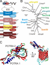
FIGURE 1.
FIGURE 1.
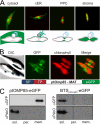
An Omp85 homolog exists in complex plastids. A, model of previously identified/suggested translocation components of each of the four membranes of complex plastids, including the Omp85 protein identified here. Secondary plastids of heterokontophytes are surrounded by four membranes. The first is in continuum with the ER (blue). The second membrane (pink) separates the PPC from the ER lumen. The intermembrane space (IMS) and the stroma (STR) are surrounded by the third (red) and fourth (brown) membranes, respectively. Nucleus-encoded plastid proteins (on top) possess a BTS at their N termini, which comprises a signal peptide (SP; dark blue) mediating cotranslational import via the Sec61 translocon. The transit peptide (TP; red) mediates translocation across the second membrane, most likely via the ER-associated protein degradation-derived SELMA complex. Stromal proteins bearing an aromatic amino acid at position +1 of the transit peptide are further translocated across the plastid envelope membranes by Omp85 (identified here) and by Tic20. B, calculation of phylogenetic relation of Omp85 proteins. Experimentally studied proteins from proteobacteria (blue), cyanobacteria (cyan), mitochondria (orange), and chloroplasts (green) and the ptOmp85 protein studied herein (yellow) are highlighted. The bootstrap values were calculated as described and are given in percent of support. C, top and side views of the homology model of ptOmp85.
FIGURE 2.
FIGURE 2.
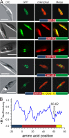
Membrane localization of the Omp85-like protein in P. tricornutum. A, distribution of GFP targeted to the cytoplasm, the ER lumen by fusion to the signal peptide of the ER luminal chaperone Bip (Bip-SP), PPC by fusion to the symbiontic Hsp70 BTS, and stroma by fusion to the AtpC BTS is shown for comparison. The model at the bottom shows the localization of GFP in the respective compartments of P. tricornutum to explain the GFP fluorescence. B, the full-length open reading frame of ptOmp85 fused to GFP was transfected into P. tricornutum. Cells expressing the construct show a green fluorescence inside the complex plastid surrounding the stroma. Shown are the differential interference contrast (DIC) image, GFP fluorescence, and chlorophyll fluorescence. Merge shows the overlay of chlorophyll and GFP fluorescence. Scale bar = 5 μm. SP, signal peptide; TP, transit peptide; MAT, mature protein; eGFP, enhanced GFP. C, P. tricornutum transfected with ptOmp85-GFP (left) or ptOmp85 BTS-GFP (BTSptOmp85-GFP; the 75 N-terminal amino acids fused to GFP) was separated into fractions representing soluble proteins (sol.), peripheral membrane proteins (per.), and integral membrane proteins (mem.). Fractions were analyzed by Western blotting with anti-GFP (top) or anti-PsbO (bottom) antibodies as indicated.
FIGURE 3.
FIGURE 3.
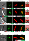
Bipartite targeting signal of ptOmp85. A, the signal peptide of 21 amino acids plus 50, 60, 62, 64, or 75 amino acids (aa; from top to bottom) fused to GFP was transfected into P. tricornutum. Shown are the differential interference contrast (DIC) image, GFP fluorescence, and chlorophyll fluorescence. Merge shows the overlay of chlorophyll and GFP fluorescence. The constructs are presented as a bar, where blue stands for the signal peptide, red for the transit peptide, yellow for the mature domain, and green for GFP. Scale bars = 5 μm. B, the hydrophobicity of the N-terminal 100 amino acids of ptOmp85 was calculated (ExPASy Proteomics Server ProtScale) in a sliding 9-amino acid window using the hydrophobicity scale established by Eisenberg et al. (53). The amino acid position was normalized to the phenylalanine at position 0. The N-terminal section of ptOmp85 is indicated as a bar diagram with the same coloring as in A. The arrow highlights the length of the sequence, where transition of GFP localization occurs.
FIGURE 4.
FIGURE 4.
Localization of ptOmp85 in the third outermost membrane. A, the self-assembling split GFP system was adapted for P. tricornutum. Either of the two fragments of GFP (S1–10 or S11) was fused to ptOmp85 (S11, first and third panels), ptMGD1 (S1–10, third panel; and S11, fourth panel), or P. tricornutum plastidal type I signal peptidase I (S1–10, third panel) or was inserted between the BTS and mature domain of ptOmp85 (S11, first and third panels). The constructs that were cotransfected are shown below each panel. Orange indicates the hydrophobic region of the signal preceding the mature domain. Processing of the samples was performed as described in the legend to Fig. 2B. On the right site of each row, a model for the positioning of the two domains of the split GFP is given in the same color code as described in the legend to Fig. 1A. MAT, mature protein. B, ptMGD1 fused to GFP as presented in the bar diagram was cotransfected into P. tricornutum, and the processing of the samples was performed as described in the legend to Fig. 2B. Scale bars = 5 μm (A and B). DIC, differential interference contrast; SP, signal peptide; TP, transit peptide; sHsp70, symbiontic Hsp70; IMS, intermembrane space; eGFP, enhanced GFP.
FIGURE 5.
FIGURE 5.
Electrophysiological properties of ptOmp85: the reversal potential. A, the reversal potential was determined by application of voltage ramps (ΔV = 10 mV/s) across bilayers (salt gradient of 250 to 20 mm KCl and 10 mm MOPS/Tris (pH 7), cis/trans). Erev is an average of 45 independent experiments. B, the charge distribution of the channel interior not considering the integrated loop is shown. Red indicates acidic regions, and blue indicates basic regions. ESP, electrostatic potential.
FIGURE 6.
FIGURE 6.
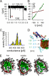
Electrophysiological properties of ptOmp85: the conductance. A, a representative current recording of a ptOmp85 bilayer at the indicated holding potential (250 mm KCl and 10 mm MOPS/Tris (pH 7), cis/trans) is shown, and the different subconductance states are marked. B, the current/voltage relationship of the two main conductance states (LARGE, green; SMALL, red) is shown. C, a histogram of subconductance states (conditions as described for A) is given. The distribution was analyzed by least-square fit to a 3-gauss equation. pS, picosiemens. D, the model for orientation of the cross-sections shown in E and the dimensions of the water-filled area are given. E, the cross-sections of the channel top, middle, and bottom are shown. The surface of the water molecules (green) and the protein (gray) and secondary structure elements are shown.

Similar articles

Cited by

References

    1. Field C. B., Behrenfeld M. J., Randerson J. T., Falkowski P. (1998) Science 281, 237–240 - PubMed
    1. Winzeler E. A. (2008) Nature. 455, 751–756 - PMC - PubMed
    1. Gould S. B., Waller R. F., McFadden G. I. (2008) Annu. Rev. Plant Biol. 59, 491–517 - PubMed
    1. Bolte K., Bullmann L., Hempel F., Bozarth A., Zauner S., Maier U. G. (2009) J. Eukaryot. Microbiol. 56, 9–15 - PubMed
    1. Hempel F., Bullmann L., Lau J., Zauner S., Maier U. G. (2009) Mol. Biol. Evol. 26, 1781–1790 - PubMed

Publication types

Substances

LinkOut - more resources