. 2010 Mar 5;285(10):7374-83.
doi: 10.1074/jbc.M109.058503. Epub 2009 Dec 30.

Nickel ions inhibit histone demethylase JMJD1A and DNA repair enzyme ABH2 by replacing the ferrous iron in the catalytic centers

Affiliations

Nickel ions inhibit histone demethylase JMJD1A and DNA repair enzyme ABH2 by replacing the ferrous iron in the catalytic centers

Haobin Chen et al. J Biol Chem. .

Erratum in

Abstract

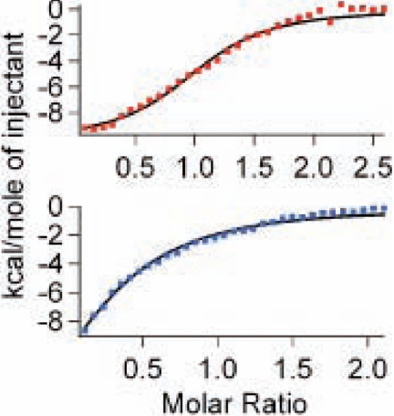
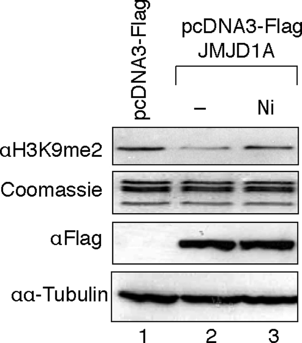
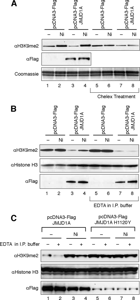
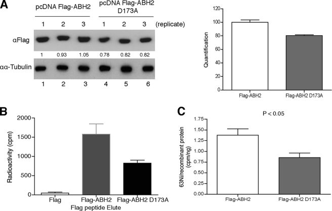
Iron- and 2-oxoglutarate-dependent dioxygenases are a diverse family of non-heme iron enzymes that catalyze various important oxidations in cells. A key structural motif of these dioxygenases is a facial triad of 2-histidines-1-carboxylate that coordinates the Fe(II) at the catalytic site. Using histone demethylase JMJD1A and DNA repair enzyme ABH2 as examples, we show that this family of dioxygenases is highly sensitive to inhibition by carcinogenic nickel ions. We find that, with iron, the 50% inhibitory concentrations of nickel (IC(50) [Ni(II)]) are 25 microm for JMJD1A and 7.5 microm for ABH2. Without iron, JMJD1A is 10 times more sensitive to nickel inhibition with an IC(50) [Ni(II)] of 2.5 microm, and approximately one molecule of Ni(II) inhibits one molecule of JMJD1A, suggesting that nickel causes inhibition by replacing the iron. Furthermore, nickel-bound JMJD1A is not reactivated by excessive iron even up to a 2 mm concentration. Using x-ray absorption spectroscopy, we demonstrate that nickel binds to the same site in ABH2 as iron, and replacement of the iron by nickel does not prevent the binding of the cofactor 2-oxoglutarate. Finally, we show that nickel ions target and inhibit JMJD1A in intact cells, and disruption of the iron-binding site decreases binding of nickel ions to ABH2 in intact cells. Together, our results reveal that the members of this dioxygenase family are specific targets for nickel ions in cells. Inhibition of these dioxygenases by nickel is likely to have widespread impacts on cells (e.g. impaired epigenetic programs and DNA repair) and may eventually lead to cancer development.


Figures

FIGURE 1.
FIGURE 1.
Nickel ion is a potent inhibitor of JMJD1A and ABH2. A, assay of FLAG-JMJD1A demethylase activity in the presence of 100 μm FeSO4 and varying concentrations of NiCl2. The gel was stained with Coomassie Blue to ensure that equal amounts of histones and FLAG-JMJD1A were added into each reaction. B, data quantification of A. Values are means ± S.D. for four replicates in two independent experiments. C, assay of ABH2 demethylase activity in the presence of 40 μm FeSO4 and varying concentrations of NiCl2. Oligo, oligonucleotide. D, data quantification of C.
FIGURE 2.
FIGURE 2.
Nickel ions inhibit JMJD1A by competing with iron ions. A, FLAG-JMJD1A was incubated with varying concentrations of NiCl2 on ice for 10 min, and its demethylase activity was subsequently measured using in vitro demethylase assay. The results of one typical experiment from three independent experiments are shown here. B, data quantification of A and comparison with Fig. 1A. Values are means ± S.D. for four replicates in two independent experiments. C, inhibition of JMJD1A by nickel ions could not be reversed by addition of excessive iron ions. Purified FLAG-JMJD1A was preincubated with 5 μm NiCl2 and was then assayed for its demethylase activity in the presence of varying concentrations of ferrous iron ions. The results of one typical experiment from two independent experiments are shown here.
FIGURE 3.
FIGURE 3.
XAS analysis of nickel binding to ABH2. A, K-edge XANES spectra for Fe-ABH2 (red), Ni-ABH2 (blue), and Ni-ABH2 + 2-oxoglutarate (green). Insets, expansions of the pre-edge XANES region showing peaks associated with 1s → 3d electronic transitions. B, unfiltered, k3-weighted EXAFS spectra (colored lines, red = Fe-ABH2, blue = Ni-ABH2 and green = Ni-ABH2 + 2-oxoglutarate) and best fits from Table 1 (black lines). FT, Fourier transform. Left, k-space spectra and fits. Right, FT-data and fits.
FIGURE 4.
FIGURE 4.
Binding isotherm of apo-ABH2 with iron (top) and nickel (bottom). The continuous line represents a fit of the data to a single-site binding model.
FIGURE 5.
FIGURE 5.
Nickel ions inhibit demethylase activity of JMJD1A in cells. 293T cells were transiently transfected with FLAG-JMJD1A expression vectors and were then exposed to 1 mm NiCl2 for 24 h. Histones were extracted and used for H3K9me2 detection by immunoblotting. The levels of overexpressed FLAG-JMJD1A were detected by immunoblotting using anti-FLAG antibody. The results of one typical experiment from three independent experiments are shown here.
FIGURE 6.
FIGURE 6.
Nickel ions directly bind to JMJD1A in cells to cause inhibition. A, assay of demethylase activity in the nuclear extracts of nickel ion-exposed cells. 293T cells were treated under the same conditions as described in Fig. 5. The nuclear extracts were isolated, and a portion of these extracts was passed through Chelex columns to eliminate any metals present in the extracts. In vitro histone demethylase assay was performed to measure histone demethylase activity of the nuclear extracts. The levels of FLAG-JMJD1A present in the nuclear extracts were assessed by immunoblotting using anti-FLAG antibody. B, assay of specific JMJD1A demethylase activity in the nickel ion-exposed cells. 293T cells were treated under the same condition as described in A. Cytoplasmic and nuclear extracts were isolated and combined. The combined lysates were subject to immunoprecipitation (I.P.) using anti-FLAG resin with or without addition of 1 mm EDTA into IP buffer. In vitro histone demethylase assay was performed to measure histone demethylase activity present in the immunoprecipitates. The same membrane was stained for histone H3 to assess the amount of histones loaded into the gel. The levels of immunoprecipitated FLAG-JMJD1A were assessed by immunoblotting using anti-FLAG antibody. C, assay of specific JMJD1A demethylase activity in cells pretreated with nickel ions. 293T cells were exposed to 150 μm NiCl2 for 3 days and were then transfected with FLAG-JMJD1A or FLAG-JMJD1A H1120Y expression vectors. Two days after the transfection, cell extracts were isolated and were subject to IP as described in B. In vitro histone demethylase assay was performed as in B. The results of one typical experiment from at least two independent experiments are shown in A–C.
FIGURE 7.
FIGURE 7.
Nickel ions bind to the iron-binding site of ABH2 in cells. A, measurement of FLAG-ABH2 and ABH2(D173A) expression levels in the nickel-treated 293T cells. 293T cells were transiently transfected with FLAG-ABH2 and FLAG-ABH2(D173A) expression vectors and then treated with 1 mm NiCl2 that contained 0.22 mCi of 63NiCl2. Expression of FLAG-ABH2 or ABH2(D173A) in cell lysates was measured by Western blot using anti-FLAG antibody. The intensity of bands was quantified using ImageJ software and marked below the graph. The quantification results were graphed on the right. B, cell lysates collected in A were subject to IP with anti-FLAG resin. The FLAG-tagged recombinant proteins were eluted with FLAG peptide, and their associated radioactivity was measured. C, 63Ni-specific radioactivity associated with FLAG-ABH2 or ABH2(D173A) was calculated. The experiment was conducted in triplicate, and values are means ± S.D. for triplicates. The difference in 63Ni-specific radioactivity between FLAG-ABH2 and FLAG-ABH2(D173A) samples is statistically significant because a two-tailed Student t test analysis gives a p value of 0.044.

Similar articles

Cited by

References

    1. International Agency for Research on Cancer (1990) Monographs on the Evaluation of Carcinogenic Risks to Humans: Chromium, Nickel and Welding, pp. 1–648, Lyon, France - PMC - PubMed
    1. Grandjean P., Andersen O., Nielsen G. D. (1988) Am. J. Ind. Med. 13, 193–209 - PubMed
    1. Costa M., Simmons-Hansen J., Bedrossian C. W., Bonura J., Caprioli R. M. (1981) Cancer Res. 41, 2868–2876 - PubMed
    1. Ke Q., Davidson T., Kluz T., Oller A., Costa M. (2007) Toxicol. Appl. Pharmacol. 219, 18–23 - PubMed
    1. Costa M., Abbracchio M. P., Simmons-Hansen J. (1981) Toxicol. Appl. Pharmacol. 60, 313–323 - PubMed

Publication types

MeSH terms