. 2010 Mar 26;285(13):9535-9544.
doi: 10.1074/jbc.M109.071332. Epub 2009 Dec 30.

Receptor interacting protein 3 suppresses vascular smooth muscle cell growth by inhibition of the phosphoinositide 3-kinase-Akt axis

Affiliations

Receptor interacting protein 3 suppresses vascular smooth muscle cell growth by inhibition of the phosphoinositide 3-kinase-Akt axis

Qian Li et al. J Biol Chem. .

Abstract

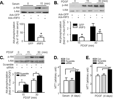
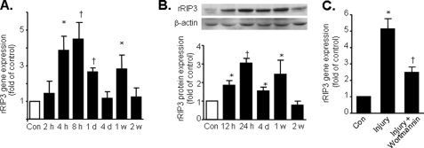
Proliferation of vascular smooth muscle cells (VSMCs) is a primary mechanism underlying cardiovascular proliferative disorders. Phosphoinositide 3-kinase (PI3K)-Akt (or protein kinase B) axis has been assigned at the center of pathways that regulate cell proliferation. Here we demonstrate that enhanced PI3K-Akt signaling by mitogenic stimulation or arterial injury profoundly elevates expression of receptor interacting protein 3 (RIP3) in primary cultured rat VSMCs and in vivo and that the up-regulation of RIP3 leads to VSMC growth arrest and apoptosis via inhibiting the PI3K-Akt signaling pathway, thereby alleviating balloon injury-induced neointimal formation. Specifically, mitogenic stimulation with platelet-derived growth factor-BB or angiotensin II leads to a profound increase in RIP3 expression, which is abolished by inhibition of PI3K or Akt, and increased PI3K-Akt signaling by expression of a constitutively active PI3K mutant also elevates RIP3 expression. Importantly, adenoviral overexpression of RIP3 not only triggers apoptosis but also causes cell cycle arrest at G(1)/G(0) phases that is associated with suppressed Akt activation. In sharp contrast, RIP3 gene silencing enhances serum- and platelet-derived growth factor-induced cell proliferation and Akt activation. In vivo adenoviral gene delivery of rat RIP3 (rRIP3) increased apoptosis and reduced VSMC proliferation, thus, effectively alleviating balloon injury-induced neointimal formation. The growth-suppressive and pro-apoptotic effects are independent of rRIP3 Ser/Thr kinase activity, because overexpression of a kinase-inactive mutant of rRIP3, similar to its wild type, is sufficient to induce growth arrest and apoptosis. These findings reveal a novel growth-suppressive action of RIP3, marking RIP3 as an important factor to prevent excessive mitogenic stimulation- or injury-induced vascular smooth muscle cells hyperplasia.


Figures

FIGURE 1.
FIGURE 1.
Up-regulation of rRIP3 in response to PDGF and Ang II treatment in primary cultured rat VSMCs. A, time courses of PDGF (10 ng/ml)- or Ang II (100 nm)-mediated increases in rRIP3 mRNA were assayed by real time PCR (*, p < 0.05; †, p < 0.01, versus control; n = 3–4). B, shown are concentration-response curves of rRIP3 mRNA to PDGF at the indicated concentrations for 4 h (†, p < 0.01 versus control; n = 3–4). C and D, time courses of PDGF (10 ng/ml)- and Ang II (100 nm)- mediated increases in rRIP3 protein level were detected by Western blots in VSMCs, respectively. In both panels, the top shows a representative Western blot, whereas the bottom illustrates the average data (*, p < 0.05; †, p < 0.01 versus control; n = 4 for each group).
FIGURE 2.
FIGURE 2.
Activation of PI3K is both necessary and sufficient for PDGF- and Ang II-mediated up-regulation of rRIP3. A, inhibition of PI3K with LY294002 (10 μm) blocks PDGF- and Ang II-induced increases in rRIP3 expression. Quiescent cells were pretreated with LY294002 (10 μm) for 30 min, then incubated with PDGF (10 ng/ml) for 4 h or Ang II (100 nm) for 6 h. rRIP3 mRNA levels were assayed by real time PCR (*, p < 0.01 versus untreated or LY294002-treated groups; n = 3 for each group). B, inhibition of PI3K with either LY294002 or wortmannin significantly attenuates a PDGF-induced increase in rRIP3 protein level. Quiescent cells were pretreated with LY294002 (10 μm) or wortmannin (50 nm) for 30 min, then incubated with PDGF (10 ng/ml) for 6 h (*, p < 0.01 versus control and cells treated with either PI3K inhibitor; †, p < 0.01 versus PDGF; n = 4 for each group). C, adenoviral expression of a constitutively activated PI3K mutant increases the rRIP3 mRNA level (*, p < 0.01 versus Adv-GFP-infected group). D, expression of the constitutively active PI3K mutant elevates rRIP3 protein abundance. The top shows typical Western blots of total (t-) or phosphorylated (p-) Akt and rRIP3, whereas the bottom illustrates the average data of rRIP3 expression. Quiescent cells were infected with Adv-GFP or Adv-CA-PI3K (100 m.o.i. for 48 h) (*, p < 0.01 versus Adv-GFP, n = 3 for each group). E and F, inhibition of Akt with a specific inhibitor (10 μm) attenuates PDGF- or Ang II-induced up-regulation of rRIP3. Cells were pretreated with the Akt inhibitor for 30 min, then treated with PDGF (10 ng/ml) for 6 h or Ang II (100 nm) for 12 h (*, p < 0.05 versus control and Akt inhibitor; †, p < 0.05 versus PDGF or Ang II; n = 4 for each group).
FIGURE 3.
FIGURE 3.
Overexpression of rRIP3 suppresses serum- and PDGF-induced VSMC growth and leads to cell cycle arrest at G0/G1 phases. A, rRIP3 protein level was assayed by Western blotting using an anti-RIP3 with total cellular proteins from cultured VSMCs infected with Adv-rRIP3 or Adv-GFP (100 m.o.i. for 24 h). B and C, shown is the inhibitory effect of rRIP3 on serum- and PDGF-stimulated VSMC growth. Quiescent VSMCs were infected with Adv-rRIP3 or Adv-GFP (100 m.o.i. for 24 h) and subsequently exposed to the serum medium (10% FBS) or PDGF (10 ng/ml). Cell numbers were determined with a hemocytometer at indicated time points (n = 5 each with triplicates; †, p < 0.01 versus Adv-GFP). D and E, overexpression of rRIP3 inhibits serum- and PDGF-stimulated VSMC proliferation assayed by MTT. Quiescent cells were infected with Adv-GFP or Adv-rRIP3 and then stimulated with 10% FBS for indicated time (†, p < 0.01 versus uninfected or Adv-GFP; n = 3). F, shown are typical examples of cell cycle distribution in VSMCs infected with Adv-GFP or Adv-rRIP3 (100 m.o.i. for 24 h) and stimulated by serum for 18 h. Then cells were stained with propidium iodide (PI) and analyzed by fluorescence-activated cell sorter (BD Biosciences FACScan). G, shown are the average data of the percent of cells in G0/G1 phases (†, p < 0.01 versus Adv-GFP; n = 3).
FIGURE 4.
FIGURE 4.
Overexpression of rRIP3 inhibits, whereas knockdown of rRIP3 augments mitogenic stimuli-induced Akt activation and cell proliferation. A and B, adenoviral overexpression of rRIP3 inhibits serum- and PDGF-induced Akt phosphorylation. t-, total; p-, phosphorylated. C, gene silencing of rRIP3 augments a PDGF-induced increase in Akt phosphorylation. The top and the bottom display, respectively, a representative Western blot, and the average data of phosphorylation of Akt at Ser-473 in cells infected with Adv-scramble and Adv-siRNA (50 m.o.i. for 72 h) and stimulated with PDGF (10 ng/ml) for the indicated times (*, p < 0.05; †, p < 0.01 as indicated; n = 3). D and E, gene silencing of rRIP3 enhances serum- and PDGF-stimulated VSMC proliferation assayed by MTT. As in panel C, quiescent VSMCs were infected with Adv-siRNA or Adv-scramble for 72 h, then stimulated with 10% FBS or PDGF (10 ng/ml) for 4 days (*, p < 0.05 versus control and Adv-scramble at day 4; n = 6–9).
FIGURE 5.
FIGURE 5.
Overexpression rRIP3 triggers apoptosis in cultured VSMCs. A and B, infection of cells with Adv-rRIP3, but not Adv-GFP, induces DNA fragmentation assayed by DNA laddering (A) and cell death ELISA (B) in cultured VSMCs. Similar results were obtained from three independent experiments for panel A. In panel B, VSMCs were harvested at 48 h after infection, and 2 × 10⁴ cells were lysed for cell death ELISA assay (*, p < 0.05; †, p < 0.01 versus Adv-GFP; n = 3). C, adenoviral expression of a constitutively active (CA) PI3K mutant blocks rRIP3-mediated apoptosis. VSMCs were infected with Adv-GFP or Adv-rRIP3 in the presence or absence of Ad-CA-PI3K (100 m.o.i. for each virus). At 48 h after infection, VSMCs were harvested, and 2 × 10⁴ cells were lysed for cell death ELISA assay (‡, p < 0.001 versus all of the other groups; n = 3). D, adenoviral overexpression of Bcl-xL protects VSMCs against rRIP3-induced apoptosis. VSMCs were infected with Adv-GFP or Adv-rRIP3 in the presence or absence of Adv-Bcl-xL (each adenovirus at m.o.i. of 100). At 48 h after infection, VSMCs were harvested, and 2 × 10⁴ cells were lysed for cell death ELISA assay (‡, p < 0.001 versus all of the other groups; n = 3). The inset shows a typical Western blot of Bcl-xL expression in cells infected by Adv-GFP or Adv-Bcl-xL at indicated m.o.i.
FIGURE 6.
FIGURE 6.
Enforced expression of a kinase-inactive rRIP3 mutant that lacks the kinase domain (rRIP3-C), similar to overexpression of wild type rRIP3, suppresses PDGF-induced VSMC growth and induces apoptosis. A, representative Western blots demonstrate adenovirus-mediated expression of rRIP3-C or its wild type counterpart (rRIP3) in primary cultured rat VSMCs. B, shown is the inhibitory effect of rRIP3-C on PDGF-stimulated VSMC growth. Quiescent VSMCs were infected with Adv-rRIP3-C, Adv-rRIP3, or Adv-GFP (all at 100 m.o.i. for 24 h) or uninfected (control) and subsequently subjected to PDGF (10 ng/ml). Cell numbers were determined with a hemocytometer at the indicated time points (n = 6 each with triplicates; *, p < 0.01 versus Adv-GFP). C, overexpression of rRIP3-C or rRIP3 inhibits PDGF-stimulated VSMC proliferation assayed by MTT. Quiescent cells were infected with Adv-GFP, Adv-rRIP3, or Adv-rRIP3-C and then stimulated with PDGF (10 ng/ml) for 4 days (*, p < 0.01 versus uninfected or Adv-GFP; n = 3). D, infection of VSMCs with Adv-rRIP3-C or Adv-rRIP3 for 24 h induces VSMC apoptosis (assayed by cell death ELISA) in a titer-dependent manner (*, p < 0.01 versus uninfected or Adv-GFP; n = 5).
FIGURE 7.
FIGURE 7.
rRIP3 is up-regulated in balloon-injured rat common carotid arteries. A, shown is a time profile of a balloon injury-induced increase in rRIP3 mRNA level (*, p < 0.05; †, p < 0.01 versus control group; n = 6). B, shown is the time profile of balloon injury-mediated increase in rRIP3 protein abundance. The top is a representative Western blot, whereas the bottom shows the average data (*, p < 0.05; †, p < 0.01 versus control group; n = 6). C, inhibition of PI3K with pretreatment of rats with wortmannin markedly suppresses a balloon injury-induced increase of rRIP3 mRNA (*, p < 0.01 versus control and wortmannin-treated groups; †, p < 0.01 versus groups in the absence of wortmannin). Rats received two intravenous injections of wortmannin (30 μg/kg) at 60 and 5 min before balloon injury. Total RNA was isolated from control and balloon-injured arteries at 8 h after surgery. Con, control.
FIGURE 8.
FIGURE 8.
Adenoviral gene transfer of rRIP3 induces VSMC apoptosis, suppresses VSMC proliferation, and inhibits neointima formation in rat carotid arteries after balloon injury. A and B, rRIP3 expression is elevated in arteries infected with Adv-rRIP3, as visualized by immunostaining with an antibody reacting with rRIP3 (A) and detected by Western blot (B). Rat common carotid arteries were dilated by balloon and infected with Adv-GFP or Adv-RIP3. On day 4 after infection, the arteries were harvested, and rRIP3 expression was detected by immunohistochemistry (A, magnification ×400) or Western blot (B). Similar results were obtained from three independent experiments in both panels. C, shown is representative TUNEL staining of cross-sections from rat carotid arteries 4 days after balloon injury in the presence of Adv-GFP or Adv-rRIP3, as indicated. Arrows indicate examples of apoptotic cells (dark brown). Original magnification, 400×. D, shown is a quantitative analysis of TUNEL positive cells (*, p < 0.01 versus Adv-GFP; n = 10 for each group). E, shown is representative PCNA staining of arterial cross-sections 1 week after surgery. F, shown are average data of PCNA staining of VSMCs (n = 6; *, p < 0.01 versus Adv-GFP). G–I, overexpression of rRIP3 in balloon-injured arteries inhibits neointimal formation. G, shown are representative histological sections of a rat left common carotid artery at 3 weeks after balloon angioplasty in the presence of Adv-GFP infection (I and III) or Adv-rRIP3 infection (II and IV) (upper panels (I and II), magnification ×100; lower panels (II and IV), magnification ×400; M, media; I, intima). H and I show, respectively, intima/media ratio and neointimal area media measured by a computer-based Image-Pro system in representative sections of the injured vessels (*, p < 0.01 versus Adv-GFP; n = 6 for each group).

Similar articles

Cited by

References

    1. Braun-Dullaeus R. C., Mann M. J., Dzau V. J. (1998) Circulation 98, 82–89 - PubMed
    1. Dzau V. J., Braun-Dullaeus R. C., Sedding D. G. (2002) Nat. Med. 8, 1249–1256 - PubMed
    1. Forrester J. S., Fishbein M., Helfant R., Fagin J. (1991) J. Am. Coll. Cardiol. 17, 758–769 - PubMed
    1. Novak K. (1998) Nat. Med. 4, 989–990 - PubMed
    1. Ferns G. A., Raines E. W., Sprugel K. H., Motani A. S., Reidy M. A., Ross R. (1991) Science 253, 1129–1132 - PubMed

Publication types

Substances

LinkOut - more resources