. 2010 Mar 5;285(10):7417-29.
doi: 10.1074/jbc.M109.053421. Epub 2009 Dec 30.

NAD+-dependent deacetylase SIRT3 regulates mitochondrial protein synthesis by deacetylation of the ribosomal protein MRPL10

Affiliations

NAD+-dependent deacetylase SIRT3 regulates mitochondrial protein synthesis by deacetylation of the ribosomal protein MRPL10

Yongjie Yang et al. J Biol Chem. .

Abstract

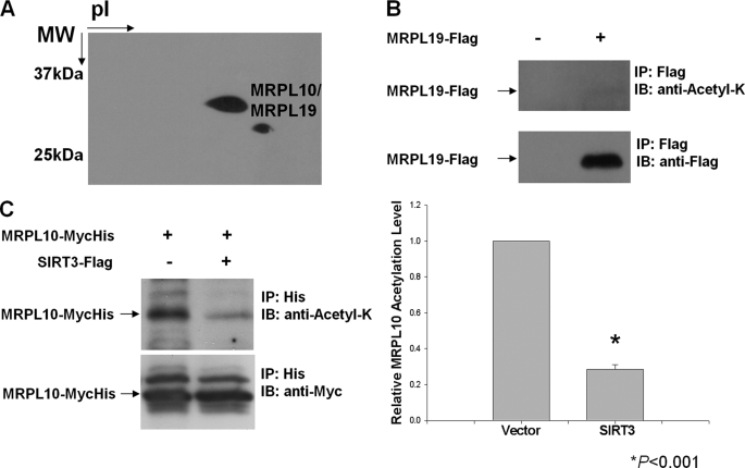
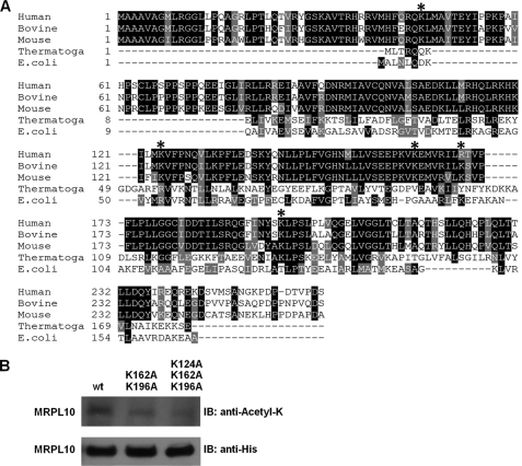
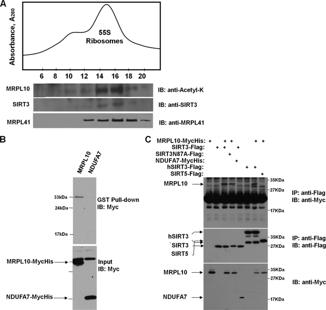
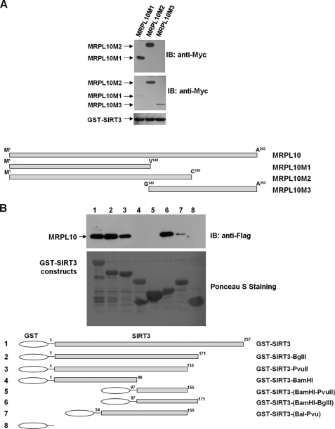
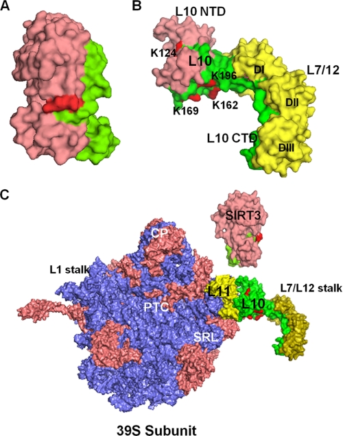
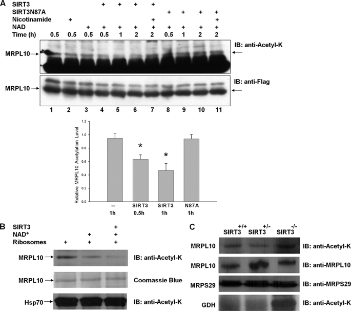
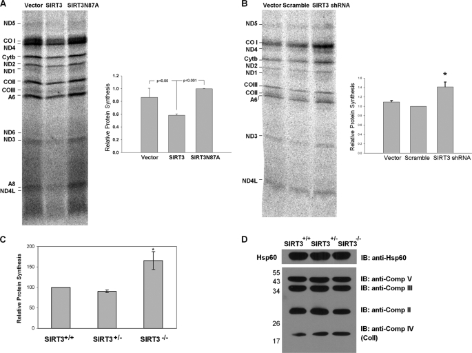
A member of the sirtuin family of NAD(+)-dependent deacetylases, SIRT3, is located in mammalian mitochondria and is important for regulation of mitochondrial metabolism, cell survival, and longevity. In this study, MRPL10 (mitochondrial ribosomal protein L10) was identified as the major acetylated protein in the mitochondrial ribosome. Ribosome-associated SIRT3 was found to be responsible for deacetylation of MRPL10 in an NAD(+)-dependent manner. We mapped the acetylated Lys residues by tandem mass spectrometry and determined the role of these residues in acetylation of MRPL10 by site-directed mutagenesis. Furthermore, we observed that the increased acetylation of MRPL10 led to an increase in translational activity of mitochondrial ribosomes in Sirt3(-/-) mice. In a similar manner, ectopic expression and knockdown of SIRT3 in C2C12 cells resulted in the suppression and enhancement of mitochondrial protein synthesis, respectively. Our findings constitute the first evidence for the regulation of mitochondrial protein synthesis by the reversible acetylation of the mitochondrial ribosome and characterize MRPL10 as a novel substrate of the NAD(+)-dependent deacetylase, SIRT3.


Figures

FIGURE 1.
FIGURE 1.
Detection of acetylated mitochondrial ribosomal proteins. Acetylated proteins found in ribosomes purified from bovine mitochondria were detected by immunoblot (IB) analysis using anti-N-acetyl lysine antibody (anti-Acetyl-K). A, approximately 1.8 A260 units of purified bovine 55 S ribosomes were separated on two-dimensional non-equilibrium pH gradient electrophoresis gels, and acetylated proteins were detected with anti-N-acetyl lysine antibody. Protein spots corresponding to the acetylated ribosomal proteins were identified by mass spectrometry. B and C, HEK293 cells were transfected with FLAG-tagged MRPL19 (B) and MycHis-tagged MRPL10 alone or together with SIRT3 (C). The tagged proteins were enriched by affinity chromatography or immunoprecipitation (IP), and MRPL10 and MRPL19 acetylation levels were detected by immunoblotting with anti-N-acetyl lysine antibody. *, p < 0.001.
FIGURE 2.
FIGURE 2.
Role of Lys124, Lys162, and Lys196 in acetylation of MRPL10. A, primary sequence alignment of MRPL10 homologs from different species. The bovine (XP_592952), human (NP_660298), and mouse (NP_080430) mitochondrial ribosomal MRPL10 proteins were aligned with Thermotoga maritimi (Thermotoga) (NP_228266) and E. coli (AAC43083) L10 proteins. *, acetylated Lys residues detected in the LC-MS/MS analysis. The alignment was created with the ClustalW program in Biology Workbench and displayed in BOXSHADE. B, approximately 20 μg of affinity-enriched lysates obtained from HeLa cells stably expressing His-tagged wild type (wt), double (K162A and K196A), and triple (K124A, K162A and K196A) mutants were loaded on 12% SDS-PAGE and probed with anti-N-acetyl lysine and anti-His tag antibodies. IB, immunoblot.
FIGURE 3.
FIGURE 3.
Interactions between SIRT3 and mitochondrial 55 S ribosomes and MRPL10. A, crude mitochondrial ribosomes were loaded on to 10–30% linear sucrose gradients to sediment 55 S ribosomes. To demonstrate the co-sedimentation of SIRT3 with the 55 S ribosome, immunoblot (IB) analyses were performed with anti-N-acetyl lysine antibody detecting the acetylated MRPL10, as well as anti-SIRT3 anti-MRPL41 antibodies, after separating 30 μl of each fraction on 12% SDS-PAGE. B, lysates prepared from HEK293 cells transfected with MycHis-tagged MRPL10 or NDUFA7 were incubated with recombinant GST-SIRT3 fusion protein immobilized on the glutathione-conjugated agarose beads. The proteins associated with the beads were analyzed by immunoblotting using anti-Myc antibody. C, HEK293 cells were transfected with MycHis-tagged MRPL10 or NDUFA7 with or without FLAG-tagged murine SIRT3, SIRT3N87A mutant, human SIRT3, or murine SIRT5, as indicated. The cell lysates were immunoprecipitated (IP) with anti-FLAG-agarose beads and detected by immunoblotting with anti-Myc or anti-FLAG antibodies.
FIGURE 4.
FIGURE 4.
Domains mediating the interaction between MRPL10 and SIRT3. A, GST-pull-down assays were performed using GST-SIRT3 fusion protein and three different in vitro translated MycHis-MRPL10 constructs, MRPL10M1, -M2, and -M3. MRPL10 proteins interacting with SIRT3 (top) and the expression levels of these MRPL10 proteins used for GST pull-down (middle) were probed using anti-Myc antibody. The GST-SIRT3 protein input (bottom) for each pull-down assay was visualized by Ponceau S staining of the membrane. B, GST-pull down assays were performed using FLAG-tagged MRPL10 expressed in HEK293 cells and various recombinant GST-SIRT3 constructs. MRPL10 associated with GST-SIRT3 was analyzed by immunoblotting using anti-FLAG antibody, and the GST fusion proteins were visualized by Ponceau S staining of the membrane. IB, immunoblot.
FIGURE 5.
FIGURE 5.
Structural model of the SIRT3 and MRPL10 interactions in the ribosomal L7/L12 stalk. A, crystal structure model of the human SIRT3 (Protein Data Bank code 3GLU) representing the MRP-L10 (green surface) interaction site and the Acecs2 peptide at the active site (red). B, structure of the L10-L7/L12 complex (Protein Data Bank code 1ZAX) from T. maritimi was used to model L7/L12 stalk in mitochondria. In the model, MRPL10 was colored pink (to represent the SIRT3 binding site) and green, and the conserved Lys residues found to be acetylated in bovine MRPL10 (shown by asterisks in Fig. 2A) were colored red. The L7/L12 dimers (DI, DII, and DIII) were colored yellow. C, models of the human SIRT3 and T. maritimi L10-L7/L12 complex were used to represent their possible interactions with 55 S ribosomes using coordinates from the E. coli 50 S subunit (Protein Data Bank code 2AW4). The 50 S ribosomal rRNAs, L10, and SIRT3 were colored blue, green, and pink, respectively. The other functional regions, such as peptidyltransferase center (PTC), central protuberance (CP), sarcin-ricin loop (SRL), L1, and L7/L12 stalks of the large subunit and ribosomal proteins (salmon) are labeled in the model. NTD, N-terminal domain; CTD, C-terminal domain. The structural model was generated by PyMol software (DeLano Scientific LLC).
FIGURE 6.
FIGURE 6.
Deacetylation of MRPL10 by NAD+-dependent deacetylase, SIRT3. A, MRPL10 protein was produced by transfecting FLAG-tagged MRPL10 into HEK293 cells and then immunoprecipitated with anti-FLAG-agarose beads. Purified MRPL10 was then incubated with or without recombinant SIRT3 or SIRT3 mutant, SIRT3N87A, together with 10 mm nicotinamide or 5 mm NAD+, as indicated. The acetylation of MRPL10 was detected by immunoblotting with anti-N-acetyl lysine. *, p < 0.005. B, in vitro deacetylation reactions of about 0.1 A260 units of 55 S bovine mitochondrial ribosomes were performed in the presence of 3 mm NAD+ and 0.2 μg of recombinant SIRT3 as labeled and detected by immunoblotting (IB) analysis probed with anti-N-acetyl lysine antibody. The arrows indicate the specific deacetylation of MRPL10 but not the acetylated Hsp70 sedimented with ribosomes by endogenous and recombinant SIRT3 in the presence of 3 mm NAD+. C, mitochondrial ribosomes were prepared as described under “Experimental Procedures” from Sirt3−/−, Sirt3+/−, and Sirt3+/+ mouse liver, and the acetylation of ribosomal protein MRPL10 was detected by immunoblot analysis probed with anti-N-acetyl lysine antibody. As a control for acetylation of glutamate dehydrogenase in the absence of SIRT3 and equal loading, immunoblots were developed with anti-N-acetyl lysine and mouse MRPL10 and MRPS29 antibodies.
FIGURE 7.
FIGURE 7.
Role of ribosome acetylation/deacetylation in mitochondrial translation. A, SIRT3 expression decreases mitochondrial protein synthesis in C2C12 cells. Mitochondrial DNA-encoded protein synthesis in C2C12 cells stably expressing vector alone, SIRT3, or SIRT3N87A. The cells were exposed to [35S]methionine in the presence of a cytosolic translation inhibitor, emetine. A representative electrophoretic pattern of newly synthesized translational products is presented. ND1, -2, -3, -4, -4L, -5, and -6, subunits of NADH dehydrogenase 1, 2, 3, 4, 4L, 5, and 6, respectively; Cytb, apocytochrome b; COI, -II, and -III, subunits I, II, and III, respectively; A6 and A8, subunits 6 and 8, respectively, of the H+-ATPase. The combined intensities of 12 mitochondrial DNA-encoded proteins from each lane were used as an indicator of mitochondrial protein synthesis. The graph is a quantification of three independent pulse-labeling experiments. B, SIRT3 knockdown increases mitochondrial protein synthesis in C2C12 cells. Mitochondrial protein synthesis in C2C12 cells stably expressing vector alone, scrambled shRNA, or SIRT3 shRNA were measured by pulse-labeling experiments as described above. The combined intensities of 12 mitochondrial DNA-encoded proteins from each lane were used as an indicator of mitochondrial protein synthesis. C, acetylated ribosomes promote mitochondrial protein synthesis in vitro. Mitochondrial ribosomes (0.05–0.1 A260 units) isolated from Sirt3+/+, Sirt3+/−, and Sirt3−/− mouse liver mitochondria were used in the poly(U)-directed in vitro translation assays described under “Experimental Procedures.” *, p < 0.05. D, immunoblotting (IB) analysis of oxidative phosphorylation complexes obtained from Sirt3+/+, Sirt3+/−, and Sirt3−/− mouse liver mitochondria. Approximately 50 μg of mitochondrial lysate from each sample was separated on 12% SDS-PAGE, and immunoblot analysis was performed with Hsp60 antibody and total oxidative phosphorylation complex antibody mixture containing Complex I subunit NDUFB8, Complex II subunit of 30 kDa, Complex III subunit Core 2, Complex IV subunit II (COII), and ATP synthase subunit α antibodies.

Similar articles

Cited by

References

    1. Kim S. C., Sprung R., Chen Y., Xu Y., Ball H., Pei J., Cheng T., Kho Y., Xiao H., Xiao L., Grishin N. V., White M., Yang X. J., Zhao Y. (2006) Mol. Cell 23, 607–618 - PubMed
    1. Jackson P. J., Harris D. A. (1986) Biochem. J. 235, 577–583 - PMC - PubMed
    1. Hallows W. C., Lee S., Denu J. M. (2006) Proc. Natl. Acad. Sci. U.S.A. 103, 10230–10235 - PMC - PubMed
    1. Dinardo M. M., Musicco C., Fracasso F., Milella F., Gadaleta M. N., Gadaleta G., Cantatore P. (2003) Biochem. Biophys. Res. Commun. 301, 187–191 - PubMed
    1. Gerhart-Hines Z., Rodgers J. T., Bare O., Lerin C., Kim S. H., Mostoslavsky R., Alt F. W., Wu Z., Puigserver P. (2007) EMBO J. 26, 1913–1923 - PMC - PubMed

Publication types

MeSH terms