Systemic propranolol reduces b-wave amplitude in the ERG and increases IGF-1 receptor phosphorylation in rat retina
- PMID: 20042659
- PMCID: PMC2868483
- DOI: 10.1167/iovs.09-4779
Systemic propranolol reduces b-wave amplitude in the ERG and increases IGF-1 receptor phosphorylation in rat retina
Abstract
Purpose: To determine whether systemic application of propranolol, a nonselective beta-adrenergic receptor antagonist, with an osmotic pump will decrease the b-wave amplitude of the electroretinogram (ERG) and increase insulin-like growth factor (IGF)-1 receptor signaling.
Methods: Young rats at 8 weeks of age were treated with saline, phentolamine, a nonselective alpha-adrenergic receptor antagonist, or propranolol, a nonselective beta-adrenergic receptor antagonist, delivered by osmotic pumps for 21 days. On the 21st day, all rats underwent electroretinographic analyses followed by collection of the retinas for protein assessment using Western blot analysis for IGF binding protein 3 (IGFBP3), IGF-1 receptor (IGF-1R), Akt, extracellular signal-related kinases 1 and 2 (ERK1/2), and vascular endothelial cell growth factor (VEGF).
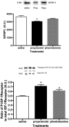
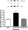
Results: Data indicate that 21 days of propranolol significantly decreased the b-wave amplitude of the ERG. The decrease in the b-wave amplitude occurred concurrently with a decrease in IGFBP3 levels and an increase in tyrosine phosphorylation of IGF-1 receptor on 1135/1136. This phosphorylation of IGF-1 receptor led to increased phosphorylation of Akt and ERK1/2. VEGF protein levels were also increased.
Conclusions: Overall, beta-adrenergic receptor antagonism produced a dysfunctional ERG, which occurred with an increase in IGF-1R phosphorylation and activation of VEGF. Systemic application of beta-adrenergic receptor antagonists may have detrimental effects on the retina.
Figures


Similar articles
-
PDGF- and insulin/IGF-1-specific distinct modes of class IA PI 3-kinase activation in normal rat retinas and RGC-5 retinal ganglion cells.Invest Ophthalmol Vis Sci. 2008 Aug;49(8):3687-98. doi: 10.1167/iovs.07-1455. Epub 2008 Apr 17. Invest Ophthalmol Vis Sci. 2008. PMID: 18421086
-
The cyclolignan picropodophyllin attenuates intimal hyperplasia after rat carotid balloon injury by blocking insulin-like growth factor-1 receptor signaling.J Vasc Surg. 2007 Jul;46(1):108-15. doi: 10.1016/j.jvs.2007.02.066. J Vasc Surg. 2007. PMID: 17606126
-
Cell-type-specific roles of IGF-1R and EGFR in mediating Zn2+-induced ERK1/2 and PKB phosphorylation.J Biol Inorg Chem. 2010 Mar;15(3):399-407. doi: 10.1007/s00775-009-0612-7. Epub 2009 Nov 28. J Biol Inorg Chem. 2010. PMID: 19946718
-
Age-associated increase in cleaved caspase 3 despite phosphorylation of IGF-1 receptor in the rat retina.J Gerontol A Biol Sci Med Sci. 2009 Nov;64(11):1154-9. doi: 10.1093/gerona/glp102. Epub 2009 Aug 20. J Gerontol A Biol Sci Med Sci. 2009. PMID: 19696229 Free PMC article.
-
Role of the adrenergic system in a mouse model of oxygen-induced retinopathy: antiangiogenic effects of beta-adrenoreceptor blockade.Invest Ophthalmol Vis Sci. 2011 Jan 5;52(1):155-70. doi: 10.1167/iovs.10-5536. Invest Ophthalmol Vis Sci. 2011. PMID: 20739470
Cited by
-
IGFBP-3 and TNF-α regulate retinal endothelial cell apoptosis.Invest Ophthalmol Vis Sci. 2013 Aug 9;54(8):5376-84. doi: 10.1167/iovs.13-12497. Invest Ophthalmol Vis Sci. 2013. PMID: 23868984 Free PMC article.
-
Topical administration of adrenergic receptor pharmaceutics and nerve growth factor.Clin Ophthalmol. 2010 Jul 21;4:605-10. doi: 10.2147/opth.s10992. Clin Ophthalmol. 2010. PMID: 20668722 Free PMC article.
-
The Beta Adrenergic Receptor Blocker Propranolol Counteracts Retinal Dysfunction in a Mouse Model of Oxygen Induced Retinopathy: Restoring the Balance between Apoptosis and Autophagy.Front Cell Neurosci. 2017 Dec 12;11:395. doi: 10.3389/fncel.2017.00395. eCollection 2017. Front Cell Neurosci. 2017. PMID: 29375312 Free PMC article.
-
Adrenergic and serotonin receptors affect retinal superoxide generation in diabetic mice: relationship to capillary degeneration and permeability.FASEB J. 2015 May;29(5):2194-204. doi: 10.1096/fj.14-269431. Epub 2015 Feb 9. FASEB J. 2015. PMID: 25667222 Free PMC article.
-
Insulin and β-adrenergic receptors inhibit retinal endothelial cell apoptosis through independent pathways.Neurochem Res. 2011 Apr;36(4):604-12. doi: 10.1007/s11064-010-0303-3. Epub 2010 Oct 30. Neurochem Res. 2011. PMID: 21053068
References
-
- Smith CP, Sharma S, Steinle JJ. Age-related changes in sympathetic neurotransmission in rat retina and choroid. Exp Eye Res 2007;84:75–81 - PubMed
-
- Holzenberger M. The GH/IGF-I axis and longevity. Eur J Endocrinol 2004;151(suppl 1):S23–S27 - PubMed
-
- Bartke A. Impact of reduced insulin-like growth factor-1/insulin signaling on aging in mammals: novel findings. Aging Cell 2008;7:285–290 - PubMed
Publication types
MeSH terms
Substances
Grants and funding
LinkOut - more resources
Full Text Sources
Molecular Biology Databases
Miscellaneous

