. 2010 Feb;176(2):800-11.
doi: 10.2353/ajpath.2010.090596. Epub 2009 Dec 30.

A mouse model of lethal synergism between influenza virus and Haemophilus influenzae

Affiliations

A mouse model of lethal synergism between influenza virus and Haemophilus influenzae

Lian Ni Lee et al. Am J Pathol. 2010 Feb.

Abstract

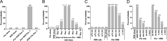
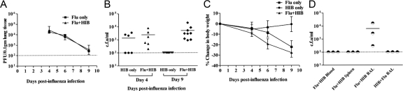
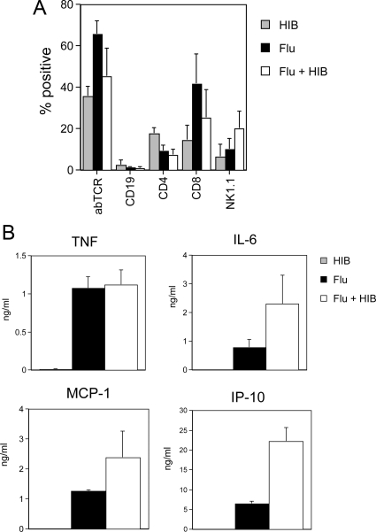
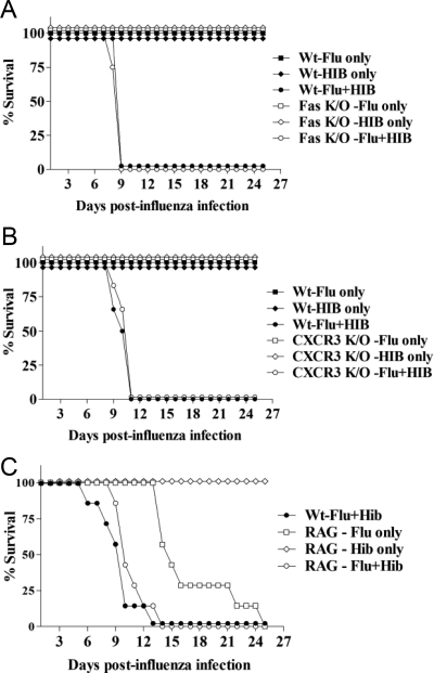
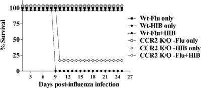
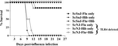
Secondary bacterial infections that follow infection with influenza virus result in considerable morbidity and mortality in young children, the elderly, and immunocompromised individuals and may also significantly increase mortality in normal healthy adults during influenza pandemics. We herein describe a mouse model for investigating the interaction between influenza virus and the bacterium Haemophilus influenzae. Sequential infection with sublethal doses of influenza and H. influenzae resulted in synergy between the two pathogens and caused mortality in immunocompetent adult wild-type mice. Lethality was dependent on the interval between administration of the bacteria and virus, and bacterial growth was prolonged in the lungs of dual-infected mice, although influenza virus titers were unaffected. Dual infection induced severe damage to the airway epithelium and confluent pneumonia, similar to that observed in victims of the 1918 global influenza pandemic. Increased bronchial epithelial cell death was observed as early as 1 day after bacterial inoculation in the dual-infected mice. Studies using knockout mice indicated that lethality occurs via a mechanism that is not dependent on Fas, CCR2, CXCR3, interleukin-6, tumor necrosis factor, or Toll-like receptor-4 and does not require T or B cells. This model suggests that infection with virulent strains of influenza may predispose even immunocompetent individuals to severe illness on secondary infection with H. influenzae by a mechanism that involves innate immunity, but does not require tumor necrosis factor, interleukin-6, or signaling via Toll-like receptor-4.


Figures

Figure 1
Figure 1
Lethal synergy between influenza virus and Hib. A: Lethal synergy between influenza virus and Hib occurs after sequential but not after simultaneous infection. Groups of four female C57BL/6 mice were infected intranasally with 30 PFU of influenza A/PR8/34 (Flu) in PBS, mock-infected with PBS or given a combination of 30 PFU flu and 105 CFU Hib in PBS. 3 days later, mice were infected with 105 CFU Hib in PBS or mock-infected with PBS. An additional group of mice was infected first with Hib and then flu 3 days later. B: Lethality is dependent on the time interval between exposure to flu and Hib. The two agents were either administered at the same time (day 0) or Hib was given 3, 4, 5, 7 or 10 days after flu. C: Effect of altering the dose of flu on lethality. Mice were infected with 105 CFU Hib, varying doses of flu or both agents 3 days apart. D: Effect of altering the dose of Hib on lethality. Mice were infected i.n. with 30 PFU flu, varying doses of Hib or both agents 3 days apart. Mice were observed for 25 days after infection. Data in each chart are shown as percent lethality. The number of mice dead/number of initial mice in each group is shown above the bar.
Figure 2
Figure 2
Sequential infection with influenza virus and Hib has no effect on viral titers but prolongs bacterial growth. A: Titers of influenza virus in the lungs. Two groups of C57BL/6 mice were infected i.n. with 30 PFU flu at day 0. At day 3, one group was also infected with 105 CFU Hib (double-infected). Lungs were collected at day 4, 6, and 9 post-flu infection from flu only (Flu) or double-infected groups. Infectious virus titers in 10% lung homogenates were determined by plaque assay on MDCK cells. Data shown are the mean titers of three mice at each time point, with the error bars depicting the SD. The experiment was performed twice. B: Bacterial counts in BAL fluid. One group of C57BL/6 mice was infected i.n. with 30 PFU flu at day 0 and then infected i.n. with 105 CFU of Hib at day 3 (Flu + Hib), together with another group of mice that were not previously infected with flu (Hib only). BAL fluid (total volume 1 ml) was obtained from the lungs at days 4 and 9 post-flu infection. Bacterial counts in BAL fluid were determined by titration on chocolate agar. Data shown are CFU bacteria/ml BAL fluid from two separate experiments. C: Weights of mice. Mice in each of the three groups were weighed at the specified time points. D: Comparison of bacterial counts in the spleen, blood and BAL of influenza and Hib co-infected mice at day 9 after infection with influenza. Mice were infected as above (Flu-Hib) or with 105 CFU of HIB followed 3 days later by 30 PFU flu (Hib-Flu). BAL was collected as described above, spleens were homogenized in 1 ml of PBS and blood was collected in heparinized tubes. Bacterial counts were determined as described above.
Figure 3
Figure 3
Lung histopathology demonstrates that mice co-infected with influenza virus and Hib show more inflammation and epithelial cell destruction than those infected by either agent alone. Groups of four mice were infected intranasally with 30 PFU influenza A/PR8 (flu), 105 CFU H. influenzae (Hib) or both agents 3 days apart (Flu + Hib). Lung samples were collected 4 and 9 days after influenza infection (1 and 6 days after Hib infection). Lung samples from uninfected mice were also collected as controls. Lungs were embedded, sectioned and hematoxylin and eosin (H&E)-stained sections were prepared and examined by microscopy. Similar results were obtained in two independent experiments. Photomicrographs of representative H&E sections are shown. A: Comparative histology of lungs from control (uninfected), Hib, Flu, or Flu + Hib-infected mice. Tissues were harvested 9 days after influenza infection (6 days after Hib infection). Lungs from mice infected with Hib alone show low levels of inflammation, while those of mice infected with flu alone contain intra-alveolar infiltrates of lymphocytes comprising a peribronchial interstitial pneumonitis with retention of alveolar spaces. In contrast, in mice infected sequentially with both agents, a pneumonitis obliterates the alveolar spaces, which are filled with neutrophils and fluid. Lymphocytes infiltrate the pulmonary interstices and extensive denudation of the bronchial epithelium is apparent. Original magnification, ×40. B: Low power comparison of lung tissue from Hib, Flu or Flu + Hib infected mice prepared as described in A. There is a progressive increase in inflammation as assessed by the degree of cellularity. Note the bronchiolar pattern in the viral pneumonia, and the focal confluent bronchopneumonia in lungs from mice infected with both agents. In comparison, a lung from a mouse infected with Hib alone shows recovery of a normal bronchial pattern and little obvious pneumonia. Original magnification, ×10. C: Histopathological features of lungs from co-infected mice at days 4 and 9 after infection with influenza (days 1 and 6 after Hib). Left: Day 4. Acute necrotizing bronchitis with neutrophils. Center: Day 4. Bronchial necrosis with apoptotic bodies and peribronchial inflammation. Right: Day 9. Erosive bronchiolitis. Original magnification, ×100. D: Histopathological scores for H&E sections of lungs from mice infected with Hib, Flu or Flu + Hib prepared as in A. Tissues were harvested 9 days after influenza infection (6 days after Hib infection). Bronchiolitis, follicular bronchitis and alveolitis were blindly scored on a scale of 0 to 3: 0, normal; 1, mild; 2, moderate; 3, severe. There was a significant difference in the scores for follicular bronchitis and alveolitis between groups of mice infected with Flu alone and Flu + Hib. *P < 0.05. **P < 0.01 (Mann-Whitney rank sum test). Scores for lungs from mice infected with Hib alone were significantly lower in all categories than those for mice infected with both flu & Hib (P < 0.001 for all categories). Data are combined from two independent experiments, with four mice per group in each experiment.
Figure 4
Figure 4
Brain histopathology of mice infected with influenza virus and Hib. Brain tissues were also collected at day 9 postinfection with influenza from the groups of infected mice described in Figure 3. Tissues were fixed, embedded in paraffin wax, sectioned and H&E-stained sections were prepared and examined by microscopy. Photomicrographs of representative H&E sections are shown. A: H&E sections of brain tissue from Hib, Flu or Flu + Hib-infected mice: Cerebral cortex with intact laminar architecture and no evidence of pia-arachnoidal inflammation. Objective magnification, ×20. B: H&E section of brain tissue (Brain) from Hib + Flu-infected mouse. Cerebellar cortex with intact molecular, Purkinje cell, and granular cell layers and no evidence of pia-arachnoidal inflammation. Objective magnification, ×20.
Figure 5
Figure 5
Cell types and cytokines in bronchoalveolar lavage from mice infected with influenza virus and Hib. Groups of three or four mice were infected intranasally with 30 PFU influenza A/PR8 (flu), 105 CFU H. influenzae (Hib) or both agents 3 days apart (Flu + Hib). Bronchoalveolar lavage samples were collected 9 days after influenza infection (6 days after Hib infection) and centrifuged to produce a cellular fraction for flow cytometric analysis of lymphocyte subsets and a cell-free supernatant for evaluation of cytokine concentrations by ELISA. A: Flow cytometric analysis of BAL cells. Cells were stained with phycoerythrin or fluorescein isothiocyanate-conjugated antibodies to the αβ T cell receptor (abTCR), CD4 or CD4 (T cells), CD19 (B cells) or NK1.1 (NK cells and NK T cells). The resulting populations were analyzed by flow cytometry using a lymphocyte gate. Mean percentage positive cells + SD are shown. B: Cytokine and chemokine concentrations in BAL. Concentrations of IL-6, TNF, MCP-1 and IP-10 were determined by ELISA. Data represent mean concentrations + SEM for two independent experiments (with the exception of MCP-1, which was from a single experiment). IL-6, TNF, IP-10, and MCP-1 were present at low to undetectable levels in BAL from mice infected with Hib alone. Levels of IP-10 were significantly increased in the BAL of coinfected mice compared with that of mice infected with flu only (P = 0.001, Mann-Whitney rank sum test).
Figure 6
Figure 6
Lethal synergy between influenza virus and Hib is not dependent on Fas, CXCR3, and T and B cells. Groups of three or four knockout mice were infected with either Flu or Hib or sequentially infected with both agents 3 days apart. As controls, groups of three or four wild-type C57BL/6 mice were also infected with both agents 3 days apart, at the same doses used for the knockout mice (B6-Flu + Hib). The mice were monitored daily and the number of days survived after flu infection was recorded. The experiment was performed twice for each knockout strain and the figures show the percentage of total mice alive in each group at each time point postinfection (N = 6–8 mice per group in total). A: Survival of Fas−/− mice after sequential infection with flu and Hib. Fas−/− mice were infected i.n. with 30 PFU of Flu (Flu only) or 105 CFU Hib (Hib only) or both agents (Flu + Hib). There was no significant difference in survival between wild-type and Fas−/− mice in any of the groups. B: Survival of CXCR3−/− mice after sequential infection with flu and Hib. CXCR3−/− mice were infected i.n. with 30 PFU Flu (Flu only) or 105 CFU Hib (Hib only) or both agents (Flu + Hib). There was no significant difference in survival between wild-type and CXCR3−/− mice in any of the groups. C: Survival of RAG−/− mice after sequential infection with Flu and Hib. RAG−/− mice were infected i.n. with 15 PFU Flu (Flu only) or 105 CFU Hib (Hib only) or both agents (RAG-Flu + Hib). The number of days survived by RAG−/− mice infected with both agents and those infected with flu only was compared in a Mann-Whitney rank sum test and was significantly different (P = 0.0006). The number of days survived by double infected RAG−/− and wild-type mice was also compared by the same test and was not significantly different.
Figure 7
Figure 7
Lethal synergy between influenza virus and Hib is not dependent on CCR2. Groups of three or four CCR2−/− mice were infected i.n. with 30 PFU Flu (Flu only) or Hib (Hib only) or both agents 3 days apart (Flu + Hib). A group of three or four wild-type C57BL/6 mice was also infected with both agents 3 days apart as a control (B6-Flu + Hib). The mice were monitored daily and the number of days survived after flu infection was recorded. The figure shows the percentage of total mice alive in each group at each time point postinfection. Combined data from two independent experiments are shown. There was no significant difference in survival between wild-type and CCR2−/− mice in any of the groups.
Figure 8
Figure 8
Lethal synergy between influenza virus and Hib is not dependent on TNF and IL-6. Groups of three or four mice lacking both TNF receptors (TNFR1R2−/−) or IL-6 (IL-6−/−) were infected i.n. with Flu or 105 CFU Hib only or both agents 3 days apart. A group of three or four wild-type C57BL/6 mice was also infected with both agents 3 days apart as control (B6-Flu + Hib). As the knockout mice were more susceptible to influenza, a lower dose of influenza was administered to all mice to minimize lethality resulting from influenza-only infection. The mice were monitored daily and the number of days survived after flu infection was recorded. The experiment was performed twice for each knockout strain. A: TNFR1R2−/− mice were infected i.n. with 20 PFU Flu (Flu only) or 105 CFU Hib (Hib only) or both agents (Flu + Hib). Wild-type control mice were also infected with both agents at the same doses. A representative experiment is shown. There was a statistically significant difference in survival of the TNFR1R2−/− mice infected with flu alone and Flu + Hib (P < 0.05, rank sum test). In contrast there was no significant difference in survival of TNFR1R2−/− and wild-type mice infected with both agents. B: Survival of IL6−/− mice after sequential infection with flu and Hib. Groups of IL6−/− mice were infected i.n. with 15 PFU Flu (Flu only) or 105 CFU Hib (Hib only) or both agents (Flu + Hib). Data combined from two independent experiments are shown. The number of days survived by IL-6−/− mice in the double-infected group was compared against the number of days survived by mice in the flu only group in a rank sum test and was significantly different (P < 0.01). The number of days survived by mice in the double-infected IL-6−/− group was also compared against double-infected wild-type C57BL/6 group by the same test and was not significantly different (P = 0.9).
Figure 9
Figure 9
Lethal synergy between influenza and Hib does not require TLR4. Groups of three C57BL/10ScNJ (TLR4−/−) mice or the wild type strain C57BL/10ScSnJ were infected i.n. with 30 PFU Flu (Flu only) or Hib (Hib only) or both agents 3 days apart (Flu + Hib). The mice were monitored daily and the number of days survived after flu infection was recorded. The figure shows the combined data from two separate experiments expressed as the percentage of total mice alive in each group at each time point postinfection (N = 6). There was no significant difference in survival between C57BL/10ScNJ and C57BL/10ScSnJ mice in any of the groups.

Comment in

Similar articles

Cited by

References

    1. Simonsen L. The global impact of influenza on morbidity and mortality. Vaccine. 1999;17(Suppl 1):S3–S10. - PubMed
    1. Thompson WW, Shay DK, Weintraub E, Brammer L, Bridges CB. Cox NJ, Fukuda K: Influenza-associated hospitalizations in the United States. JAMA. 2004;292:1333–1340. - PubMed
    1. Wong CM, Chan KP, Hedley AJ, Peiris JS. Influenza-associated mortality in Hong Kong. Clin Infect Dis. 2004;39:1611–1617. - PubMed
    1. Yap FH, Ho PL, Lam KF, Chan PK, Cheng YH, Peiris JS. Excess hospital admissions for pneumonia, chronic obstructive pulmonary disease, and heart failure during influenza seasons in Hong Kong. J Med Virol. 2004;73:617–623. - PubMed
    1. Sethi S. Bacterial pneumonia. Managing a deadly complication of influenza in older adults with comorbid disease. Geriatrics. 2002;57:56–61. - PubMed

Publication types

MeSH terms