

The immunoconjugate "icon" targets aberrantly expressed endothelial tissue factor causing regression of endometriosis
- PMID: 20042667
- PMCID: PMC2808107
- DOI: 10.2353/ajpath.2010.090757
The immunoconjugate "icon" targets aberrantly expressed endothelial tissue factor causing regression of endometriosis
Abstract
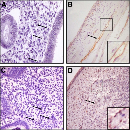
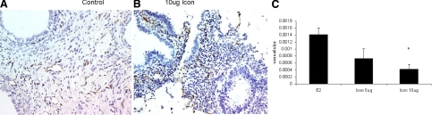
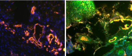
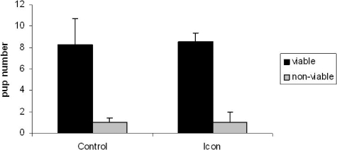
Endometriosis is a major cause of chronic pain, infertility, medical and surgical interventions, and health care expenditures. Tissue factor (TF), the primary initiator of coagulation and a modulator of angiogenesis, is not normally expressed by the endothelium; however, prior studies have demonstrated that both blood vessels in solid tumors and choroidal tissue in macular degeneration express endothelial TF. The present study describes the anomalous expression of TF by endothelial cells in endometriotic lesions. The immunoconjugate molecule (Icon), which binds with high affinity and specificity to this aberrant endothelial TF, has been shown to induce a cytolytic immune response that eradicates tumor and choroidal blood vessels. Using an athymic mouse model of endometriosis, we now report that Icon largely destroys endometriotic implants by vascular disruption without apparent toxicity, reduced fertility, or subsequent teratogenic effects. Unlike antiangiogenic treatments that can only target developing angiogenesis, Icon eliminates pre-existing pathological vessels. Thus, Icon could serve as a novel, nontoxic, fertility-preserving, and effective treatment for endometriosis.
Figures

References
-
- Giudice LC, Kao LC. Endometriosis. Lancet. 2004;364:1789–1799. - PubMed
-
- Sharpe-Timms KL, Young SL. Understanding endometriosis is the key to successful therapeutic management. Fertil Steril. 2004;81:1201–1203. - PubMed
-
- Taylor RN, Lebovic DI, Mueller MD. Angiogenic factors in endometriosis. Ann NY Acad Sci. 2002;955:89–100. discussion 118:396–406. - PubMed
-
- Nezhat C, Crowgey SR, Garrison CP. Surgical treatment of endometriosis via laser laparoscopy and videolaseroscopy. Contrib Gynecol Obstet. 1987;16:303–312. - PubMed
-
- Surrey ES, Schoolcraft WB. Management of endometriosis-associated infertility. Obstet Gynecol Clin North Am. 2003;30:193–208. - PubMed
Publication types
MeSH terms
Substances
Grants and funding
LinkOut - more resources
Full Text Sources
Other Literature Sources
Medical
Miscellaneous
