. 2010 Feb;176(2):861-9.
doi: 10.2353/ajpath.2010.090532. Epub 2009 Dec 30.

Evidence for proteotoxicity in beta cells in type 2 diabetes: toxic islet amyloid polypeptide oligomers form intracellularly in the secretory pathway

Affiliations

Evidence for proteotoxicity in beta cells in type 2 diabetes: toxic islet amyloid polypeptide oligomers form intracellularly in the secretory pathway

Tatyana Gurlo et al. Am J Pathol. 2010 Feb.

Abstract

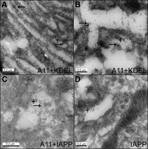
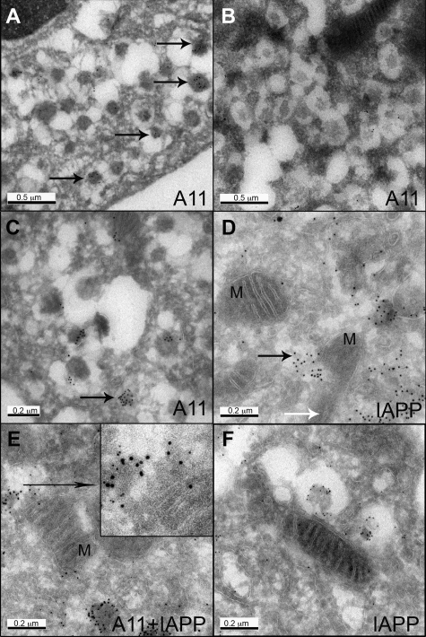
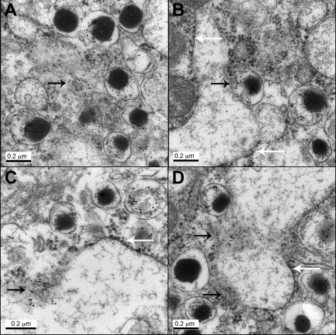
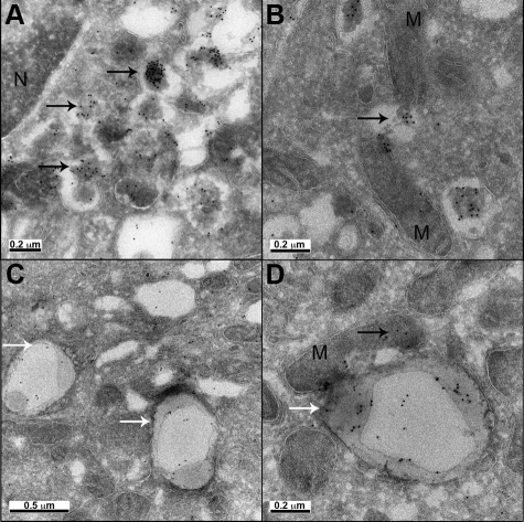
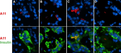
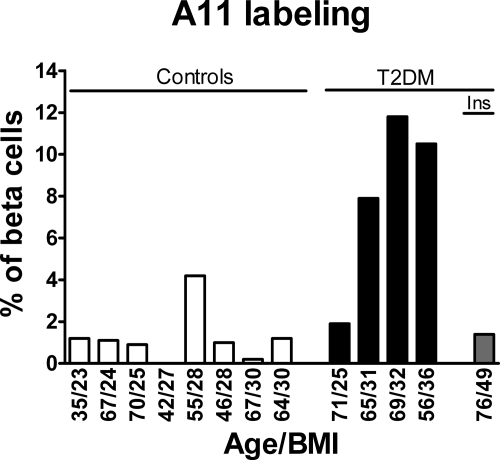
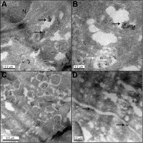
The islet in type 2 diabetes mellitus (T2DM) is characterized by a deficit in beta cells and islet amyloid derived from islet amyloid polypeptide (IAPP), a protein co-expressed with insulin by beta cells. It is increasingly appreciated that the toxic form of amyloidogenic proteins is not amyloid but smaller membrane-permeant oligomers. Using an antibody specific for toxic oligomers and cryo-immunogold labeling in human IAPP transgenic mice, human insulinoma and pancreas from humans with and without T2DM, we sought to establish the abundance and sites of formation of IAPP toxic oligomers. We conclude that IAPP toxic oligomers are formed intracellularly within the secretory pathway in T2DM. Most striking, IAPP toxic oligomers appear to disrupt membranes of the secretory pathway, and then when adjacent to mitochondria, disrupt mitochondrial membranes. Toxic oligomer-induced secretory pathway and mitochondrial membrane disruption is a novel mechanism to account for cellular dysfunction and apoptosis in T2DM.


Figures

Figure 1
Figure 1
Oligomer-specific immunoreactivity was found in association with the ER membrane and in the distended ER lumen (A, B), and in the Golgi system (C, D). Sections of islets from hIAPP transgenic mice were labeled for oligomers (A11) and ER marker KDEL (A, B; 10 nm gold and 5 nm gold, respectively); co-labeled for oligomers (A11) and IAPP (C, 5 nm and 10 nm gold respectively); labeled for IAPP (D; 10 nm gold). Long arrows point to oligomers, short arrows point to KDEL (A, B) or IAPP (C, D). Original magnification: ×100,000 (A, C, D) and ×150,000 (B).
Figure 2
Figure 2
Oligomer immunoreactivity (A11, 10 nm gold) was found in the secretory vesicles (A, arrows) and diffuse aggregates (C, arrow) in β cells from hIAPP transgenic mice, but only occasional weak labeling in β cells from rIAPP transgenic mice (B). D, E: IAPP and oligomer labeled aggregates were found adjacent to mitochondria, and mitochondria (M) integrity appeared to be compromised (black arrow points to the aggregates penetrating mitochondria; white arrow points to the remnant of cristae in damaged mitochondria). F: For comparison, despite the presence of an immediately adjacent vesicle labeled for IAPP, the mitochondrion is intact in a β cell from a rIAPP transgenic mouse (5 nm gold). Original magnification: ×60,000 (A, B); ×100,000 (C, F); ×120,000 (D, E).
Figure 3
Figure 3
Immunogold labeling for IAPP in β cells from hIAPP transgenic mice (regular electron microscopy). A: Immunoreactivity was found in secretory vesicles and diffuse aggregates in the cytoplasm (arrow). B: Secretory vesicle appears to discharge IAPP (black arrows) into the ER lumen. C, D: Diffuse aggregates were found in association with ER membrane that lost its integrity (black arrow). White arrows point to the ribosomes at the ER membrane. Original magnification: ×100,000 (A, B, D), and ×120,000 (C).
Figure 4
Figure 4
Electron micrographs of human insulinoma cells expressing IAPP. A: Oligomer immunoreactivity (10 nm gold) was detected in perinuclear area in vesicle-like structures and aggregates (N, nucleus). B: Some oligomer aggregates appear to compromise mitochondria (M) integrity. C, D: Oligomer labeling was detected in vacuoles located in membrane rich areas, which appear to be autophagosomes. Black arrows point to oligomers (A, B, D); white arrows point to autophagosomes (C, D). Original magnification: ×60,000 (C), ×80,000 (A), ×100,000 (B, D).
Figure 5
Figure 5
A11 immunoreactivity was detected in β cells from T2DM cases in different patterns. A: Spotted (a, image from case #4). B: Perinuclear (image from case #2). C: Cytoplasmic (image from insulinoma case with prior T2DM). D: In some occasions, staining was detected inside the amyloid deposits (image from insulinoma case with prior T2DM). Nuclei were visualized with DAPI.
Figure 6
Figure 6
Oligomer labeling in human β cells is more frequent in T2DM cases then in controls, and tends to increase with obesity in T2DM cases. Ins, case with insulinoma (text and Supplemental Table S1 at http:/ajp.amjpathol.org for details).
Figure 7
Figure 7
Electron micrographs of human β cells from T2DM case (case #3) labeled with A11 antibody (10 nm gold). A: Oligomer immunoreactivity was detected in perinuclear area in aggregates associated with ER (N - nucleus); B: in Golgi and abnormal diffuse aggregates in poorly defined structures (black and white arrows, respectively); C: in secretory vesicles, and D in association with plasma membrane. Arrows point to oligomer aggregates. Original magnification 1:60,000 (C), 1:80,000 (A, D), 1:100,000 (B).

Similar articles

Cited by

References

    1. Butler AE, Janson J, Bonner-Weir S, Ritzel R, Rizza RA, Butler PC. Beta-cell deficit and increased beta-cell apoptosis in humans with type 2 diabetes. Diabetes. 2003;52:102–110. - PubMed
    1. Weyer C, Bogardus C, Mott DM, Pratley RE. The natural history of insulin secretory dysfunction and insulin resistance in the pathogenesis of type 2 diabetes mellitus. J Clin Invest. 1999;104:787–794. - PMC - PubMed
    1. Haataja L, Gurlo T, Huang CJ, Butler PC. Islet amyloid in type 2 diabetes, and the toxic oligomer hypothesis. Endocr Rev. 2008;29:303–316. - PMC - PubMed
    1. Ohsawa H, Kanatsuka A, Yamaguchi T, Makino H, Yoshida S. Islet amyloid polypeptide inhibits glucose-stimulated insulin secretion from isolated rat pancreatic islets. Biochem Biophys Res Commun. 1989;160:961–967. - PubMed
    1. Butler PC, Chou J, Carter WB, Wang YN, Bu BH, Chang D, Chang JK, Rizza RA. Effects of meal ingestion on plasma amylin concentration in NIDDM and nondiabetic humans. Diabetes. 1990;39:752–756. - PubMed

Publication types

MeSH terms