. 2010 Feb;176(2):903-13.
doi: 10.2353/ajpath.2010.090495. Epub 2009 Dec 30.

Expression of the embryonic stem cell transcription factor SOX2 in human skin: relevance to melanocyte and merkel cell biology

Affiliations

Expression of the embryonic stem cell transcription factor SOX2 in human skin: relevance to melanocyte and merkel cell biology

Alvaro C Laga et al. Am J Pathol. 2010 Feb.

Abstract

SOX2 is a gene located on chromosome 3q26.33 that encodes a transcription factor important to maintenance of embryonic neural crest stem cell pluripotency. We have identified rare SOX2-immunoreactive cells in normal human skin at or near the established stem cell niches. Three subsets of SOX2-positive cells were defined in these regions: those expressing only SOX2 and those that co-expressed SOX2 and either CK20 or microphthalmia-associated transcription factor, which are consistent with dichotomous differentiation of SOX2-expressing precursors along neuroendocrine (Merkel cell) or melanocytic lines, respectively. Examination of Merkel cell carcinomas confirmed nuclear SOX2 expression in this tumor type. In human patient melanoma, strong nuclear expression of SOX2 was noted in a subset of tumors, and the ability to detect SOX2 in lesional cells significantly correlated with primary tumor thickness in a survey cohort. To assess the potential role of SOX2 in melanoma growth, an in vivo tumorigenesis assay was used. Whereas SOX2 knockdown failed to influence proliferation of cultured melanoma cells in vitro, tumor xenografts generated with the SOX2-knockdown cell line showed significant decrease in mean tumor volume as compared with controls. In aggregate, these findings suggest that SOX2 is a novel biomarker for subpopulations of normal skin cells that reside in established stem cell niches and that might relate to Merkel cell and melanocyte ontogeny and tumorigenesis.


Figures
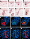
Figure 1
Figure 1
SOX2 immunoreactivity in embryonic and fetal human skin. A: At 8 to 10 weeks of EGA, scattered SOX2-immunoreactive cells are present in the dermis. Some of these cells are intimately associated with vascular channels. B: In the early developing follicles typical of 11 to 12 weeks EGA, SOX2-positive cells are evident within the primary hair germ. C and D: Follicles typical of week 13 to 14 EGA show SOX2 positivity in mesenchymal cells forming the rudiment of follicular papillae beneath the hair germ. E: At the hair peg embryonic stage (∼15 weeks EGA), SOX2 labels most cells in follicular papillae, and some SOX2-positive cells appear to migrate into the follicular adventitial sheath enclosing the forming follicle. F and G: During the bulbous hair peg stage (15 to 19 weeks EGA), SOX2-positive cells in the follicular papillae become increasingly prominent and spherical in configuration. Occasional SOX2-positive cells are apparent within the infundibular epithelium of some hair follicles (F, inset). H: In differentiated lanugo follicles (> 19 weeks EGA), cells forming follicular papillae characteristically surrounded in part by follicular matrix epithelium, and contiguous cells extending within the adventitial sheath of the hair bulb are stained by SOX2. I–K: Follicular papilla cells show immunoreactivity for SOX2 (red), and a subpopulation of SOX2-positive cells co-express nestin (green); co-expression elsewhere in the follicle was not observed. L–N: Another differentiated follicle exhibits mutually exclusive SOX2-positivity (red) in the follicular papilla and SOX10 positivity (green) within melanocytes of the hair bulb. Magnification: A: ×100; B–E: ×400; F: ×100 (inset ×400); G: ×200; H: ×400; I–N: ×1000; blue = DAPI stain.
Figure 2
Figure 2
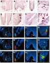
SOX2 immunoreactivity in adult human skin. A–C: SOX2-immunoreactive cells localize to the basal cells of the epidermis, principally at the tips of rete ridges. In contrast, the early embryonic skin devoid of rete ridges shows occasional SOX2-positive cells in the stratum germinativum and in the dermis (A, inset). D: Buccal mucosa, also normally devoid of rete ridges, shows SOX2-positive cells within the basal layer and superficial submucosa. E and F: In adult hair follicles, SOX2-positive cells are identified in the bulge region that approximates the insertion site of the arrector pili muscle. G: During telogen, numerous SOX2-positive cells are present in the condensed mesenchyme that surrounds the residual permanent portion of the hair follicle. H: These cells are intimately associated with persistent intraepithelial SOX2-positive cells. I–K: Double-label immunofluorescence of adult human skin shows mutually exclusive SOX2 (red, arrow) and MITF (green, arrow) nuclear positivity in rete-associated cells. L–N: Rare rete-associated cells, however, co-express both biomarkers (rete tip contour defined by dotted line). O–Q: SOX2 (red) and CK20 (green) immunoreactive cells are identified at or near rete ridges (note SOX2-only cell [arrow]). R: CK20 (green) cytoplasmic positivity is variably expressed in SOX2 (red)/CK20 double-labeled cells, while (S) other CK20-positive cells fail to express SOX2. T: Triple label showing rete-associated cluster of SOX2-only cell (red nuclear), MITF-only cell (green nuclear reactivity in adjacent cell), and SOX2 and CK20 co-expression (red nuclear and green cytoplasmic). Magnification: A and inset: ×100; B: ×400; C: ×600; D: ×400; E: ×100; F: ×600; G: ×100; H: ×600; I–K: ×1000; L–N: ×10000; O–Q: ×1000; R–T: ×6000; blue = DAPI stain.
Figure 3
Figure 3
SOX2 immunoreactivity in Merkel cell carcinoma. A: Primary cutaneous neuroendocrine carcinoma showing diffuse positivity (B) for SOX2. C–D: The intensity of nuclear immunoreactivity typically varied among cells that infiltrated the dermal connective tissue. E–H: Merkel cell carcinoma with squamous differentiation exhibiting SOX2 immunoreactivity in the neuroendocrine but not the squamous component (G). Note zonal positivity among cells forming the neuroendocrine component (H). I: The neuroendocrine component co-expressed SOX2 (brown) and CK20 (dark blue-black perinuclear dot-like pattern), with the squamous component negative for both markers. J–K: High magnification of markedly enlarged neoplastic squamous cell devoid of SOX2 or CK20 reactivity. L: Merkel cell carcinoma with striking morphological similarity to small cell/envoid melanoma with co-expression of SOX2 and CK20. M: SOX2/CK20 immunoperoxidase double labeling disclosing a small focus of CK20-positive tumor cells within a vascular lumen. Note the strong nuclear immunoreactivity for SOX2 (brown) in comparison with the less conspicuous CK20-positivity in a dot-like pattern (dark blue-black). Magnification: A and B: ×100; C and D: ×400; E: ×100; F and G: ×200; H: ×400; I: ×200; J and K: ×400; L: ×100; M: ×400.
Figure 4
Figure 4
SOX2 immunoreactivity in human melanoma. A: Primary nodular melanoma showing B and C: SOX2 immunoreactivity accentuated at the deep and peripheral edges of the tumor near the interface with underlying stroma. Occasional SOX2-positive cells were observed in the bulge regions of surrounding hair follicles (B, inset). The majority of nevi were not immunoreactive with SOX2 (C, inset). D and E: Cells of desmoplastic/spindle melanoma showing SOX2 reactivity in neurotropic foci. F and G: SOX2 reactivity in small cell metastatic melanoma. H–K: Bimorphic epithelioid and spindle cell components of primary melanoma vertical growth showing relative SOX2-negativity in the former (H and I) and positivity in the latter (J and K). L: SOX2-positive tumor cells in regions of spindle cell vertical growth tend to associate with vessels (arrows) in peritumoral stroma. M: Double label showing SOX2-positive melanoma cells (brown) associated with numerous stromal CD31-positive vessels (dark blue-black). N and O: Double label immunofluorescence further confirming intimate association of SOX2-positive cells (red) with CD31-positive blood vessels (green, blue = DAPI stain). Magnification: A and B: ×40; C ×100 (B and C, insets: ×100); D and E: ×100; F and G: ×400; H–K: ×100; L: ×400; M: ×100; N: ×200; O: ×400.
Figure 5
Figure 5
SOX2 knockdown in A2058 melanoma cells inhibits tumor growth in vivo but not in vitro. A: SOX2 knockdown by lentiviral shRNA. Stable cell lines expressing lentiviral shRNA for human SOX2 and non-target control were subjected to Western blotting using β-actin as internal loading control. Near 70% Sox2 knockdown efficiency was achieved as determined by densitometry. B: Cell growth under conventional monolayer culture conditions. Both SOX2 knockdown clone C (Sox2 KD cl. C) and control cells exhibit comparable growth rates in vitro. C: Tumorigenicity in vivo. SOX2 KD significantly inhibits tumorigenicity of A2058 melanoma cells, compared with the control. D: Immunohistochemistry shows that the tumor xenografts derived from SOX2 KD cl. C cells are devoid of SOX2 expression, while those of control cells retain constitutive SOX2 expression.

Similar articles

Cited by

References

    1. Bittner M, Meltzer P, Chen Y, Jiang Y, Seftor E, Hendrix M, Radmacher M, Simon Rm, Yakhini Z, Ben-Dor A, Sampas N, Dougherty E, Wang E, Marincola F, Gooden C, Lueders J, Glatfelter A, Pollock P, Carpten J, Gillanders E, Leja D, Dietrich K, Beaudry C, Berens M, Alberts D, Sondak V. Molecular classification of cutaneous malignant melanoma by gene expression profiling. Nature. 2000;406:536–540. - PubMed
    1. Hendrix MJ, Seftor EA, Hess AR, Seftor RE. Molecular plasticity of human melanoma cells. Oncogene. 2003;22:3070–3075. - PubMed
    1. Simpson AJ, Caballero OL, Jungbulth A, Chen YT, Old LJ. Cancer/testis antigens, gametogenesis, and cancer. Nat Rev Cancer. 2005;5:615–625. - PubMed
    1. Rothhammer T, Wild PJ, Meyer S, Bataille F, Pauer F, Pauer A, Klinkhammer-Schalke M, Hein R, Hofstaedter F, Bosserhoff AK. Bone morphogenic protein 7 (BMP7) expression is a potential novel prognostic marker for recurrence in patients with primary melanoma. Cancer Biomark. 2007;3:111–117. - PubMed
    1. Hsu MY, Rovinsky SA, Lai CY, Qasem S, Liu X, How J, Engelhardt JF, Murphy GF. Aggressive melanoma cells escape from BMP7-mediated autocrine growth inhibition through coordinated Noggin upregulation. Lab Invest. 2008;88:842–855. - PMC - PubMed

Publication types

MeSH terms