. 2010 Feb;176(2):549-55.
doi: 10.2353/ajpath.2010.090722. Epub 2009 Dec 30.

Ectopic T-cell specificity and absence of perforin and granzyme B alleviate neural damage in oligodendrocyte mutant mice

Affiliations

Ectopic T-cell specificity and absence of perforin and granzyme B alleviate neural damage in oligodendrocyte mutant mice

Antje Kroner et al. Am J Pathol. 2010 Feb.

Erratum in

  • Am J Pathol. 2010 Oct;177(4):2145

Abstract

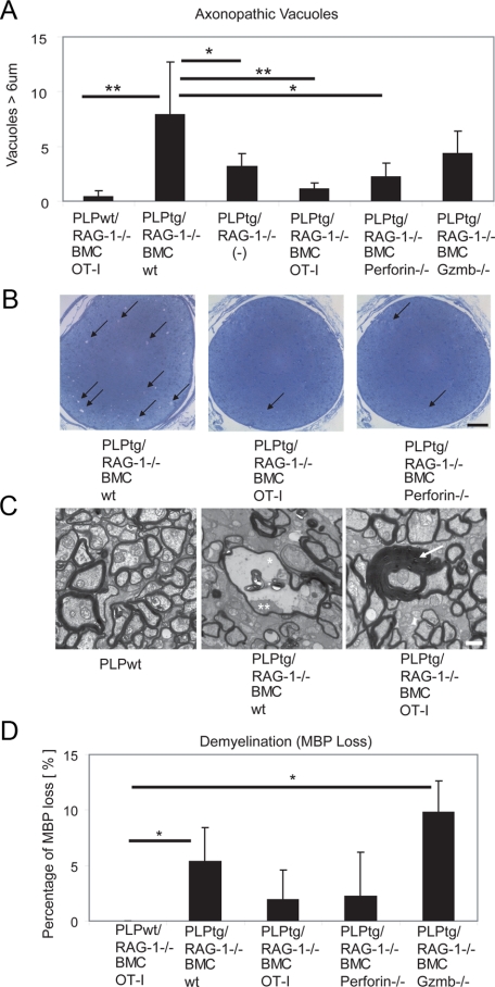
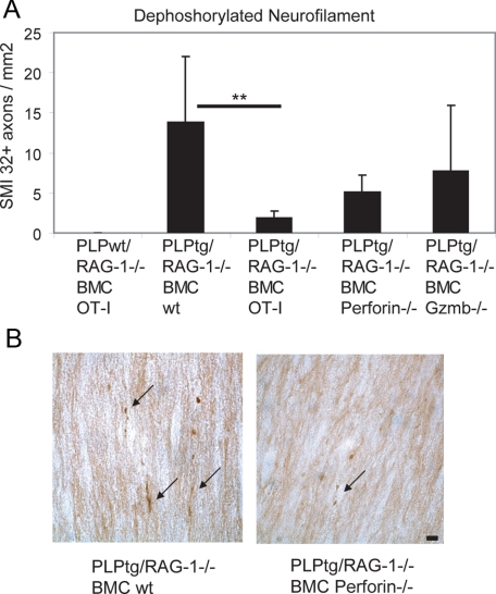
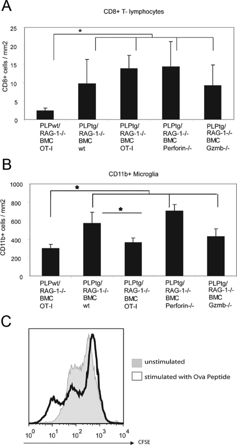
In transgenic mice overexpressing the major myelin protein of the central nervous system, proteolipid protein, CD8+ T-lymphocytes mediate the primarily genetically caused myelin and axon damage. In the present study, we investigated the cellular and molecular mechanisms underlying this immune-related neural injury. At first, we investigated whether T-cell receptors (TCRs) are involved in these processes. For this purpose, we transferred bone marrow from mutants carrying TCRs with an ectopic specificity to ovalbumin into myelin mutant mice that also lacked normal intrinsic T-cells. T-lymphocytes with ovalbumin-specific TCRs entered the mutant central nervous system to a similar extent as T-lymphocytes from wild-type mice. However, as revealed by histology, electron microscopy and axon- and myelin-related immunocytochemistry, these T-cells did not cause neural damage in the myelin mutants, reflecting the need for specific antigen recognition by cytotoxic CD8+ T-cells. By chimerization with bone marrow from perforin- or granzyme B (Gzmb)-deficient mice, we demonstrated that absence of these cytotoxic molecules resulted in reduced neural damage in myelin mutant mice. Our study strongly suggests that pathogenetically relevant immune reactions in proteolipid protein-overexpressing mice are TCR-dependent and mediated by the classical components of CD8+ T-cell cytotoxicity, perforin, and Gzmb. These findings have high relevance with regard to our understanding of the pathogenesis of disorders primarily caused by genetically mediated oligodendropathy.


Figures

Figure 1
Figure 1
Neural damage in PLPtg mice depends on an intact TCR repertoire and molecules typical for cytotoxic T-lymphocytes. A: Quantification of periaxonal vacuoles in semithin sections of optic nerves from PLPwt mice and PLPtg mutants with normal or molecularly altered T-lymphocytes. Note reduction in vacuole numbers when T-lymphocytes lack relevant TCR (OT-I) or the cytotoxic molecules perforin and Gzmb. B: Representative semithin sections from PLPtg mutants with wild-type immune system, with irrelevant TCR (OT-I) and with lymphocytes lacking perforin (perforin−/−). Note reduced vacuole numbers when T-lymphocytes lack relevant TCR (OT-I) or the cytotoxic molecule perforin. Arrows point to periaxonal vacuoles. Scale bar = 50 μm. C: Representative electron micrographs from optic nerves of PLPwt mice, PLPtg mutants with wild-type immune system (PLPtg/RAG-1−/− BMCwt) and of PLPtg mutants with irrelevant TCR (PLPtg/RAG-1−/− OT-I). Note normal appearance of myelin in the PLPwt mice (left) and occurrence of myelin vacuoles in PLPtg mutants with wild-type immune system (middle; asterisk). Remnant of an axon is indicated by a double asterisk. In PLPtg mutants with irrelevant TCR (right), demyelination and formation of vacuoles is substantially reduced. Occasionally, formation of redundant myelin loops (arrow) is seen. Scale bar = 1 μm. D: Reduction in MBP immunoreactivity in optic nerves from PLPtg mutants with normal or molecularly altered T-lymphocytes indicative of demyelination. Note that loss of MBP is minimized in the absence of relevant TCR or perforin, whereas absence of Gzmb apparently does not reduce demyelination. PLPwt mice do not show demyelination. *P < 0.05, **P ≤ 0.01, n = 3 to 6 per group.
Figure 2
Figure 2
Axonal damage in optic nerves from PLPwt and PLPtg mutants with normal or molecularly altered T-lymphocytes. A: Quantification of dephosphorylated neurofilament (SMI32) in optic nerve longitudinal sections. Note reduction of numbers of profiles with dephosphorylated neurofilaments in PLPtg mutants with irrelevant TCR (OT-I) or in the absence of cytotoxic molecules, while PLPwt mice do not display SMI32+ profiles. **P ≤ 0.01. B: Representative examples of SMI32+ profiles (arrows) in optic nerves of PLPtg mutants with wild-type immune system or in the absence of perforin. Note reduced SMI32+ profiles in the latter. Scale bar = 10 μm.
Figure 3
Figure 3
Number of immune cells in optic nerves from PLPwt and PLPtg mutants with normal or molecularly altered T-lymphocytes. A: As shown previously, CD8+T-lymphocytes are more frequent in PLPtg mice compared with PLPwt mice. Note that in all types of PLPtg mutants, numbers of CD8+T-lymphocytes are similar, reflecting their unchanged ability to enter the mutant CNS. B: Numbers of CD11b+ microglial cells are higher in PLPtg as compared with PLPwt mice. PLPtg mutants with irrelevant TCR show a reduced number of microglial cells. C: Histogram overlay of carboxyfluorescein succinimidyl ester-labeled splenocytes stimulated with Ova peptide (black) or unstimulated (gray). Reduction of carboxyfluorescein succinimidyl ester intensity reflects proliferation. *P < 0.05, n = 3 to 6 per group.

Similar articles

Cited by

References

    1. Ip CW, Kroner A, Fischer S, Berghoff M, Kobsar I, Mäurer M, Martini R. Role of immune cells in animal models for inherited peripheral neuropathies. Neuromol Med. 2006;8:175–189. - PubMed
    1. Ip CW, Kroner A, Bendszus M, Leder C, Kobsar I, Fischer S, Wiendl H, Nave KA, Martini R. Immune cells contribute to myelin degeneration and axonopathic changes in mice overexpressing proteolipid protein in oligodendrocytes. J Neurosci. 2006;26:8206–8216. - PMC - PubMed
    1. Ip CW, Kroner A, Crocker PR, Nave KA, Martini R. Sialoadhesin deficiency ameliorates myelin degeneration and axonopathic changes in the CNS of PLP overexpressing mice. Neurobiol Dis. 2007;25:105–111. - PubMed
    1. Leder C, Schwab N, Ip CW, Kroner A, Nave KA, Dornmair K, Martini R, Wiendl H. Clonal expansions of pathogenic CD8+ effector cells in the CNS of myelin mutant mice. Mol Cell Neurosci. 2007;36:416–424. - PubMed
    1. Kassmann CM, Lappe-Siefke C, Baes M, Brugger B, Mildner A, Werner HB, Natt O, Michaelis T, Prinz M, Frahm J, Nave KA. Axonal loss and neuroinflammation caused by peroxisome-deficient oligodendrocytes. Nat Genet. 2007;39:969–976. - PubMed

Publication types

MeSH terms