. 2010 Feb;176(2):914-25.
doi: 10.2353/ajpath.2010.090744. Epub 2009 Dec 30.

Blockade of autoantibody-initiated tissue damage by using recombinant fab antibody fragments against pathogenic autoantigen

Affiliations

Blockade of autoantibody-initiated tissue damage by using recombinant fab antibody fragments against pathogenic autoantigen

Gang Wang et al. Am J Pathol. 2010 Feb.

Abstract

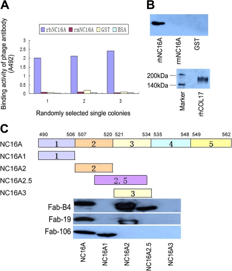
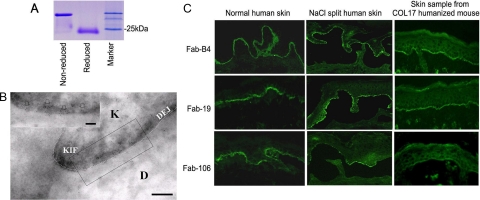
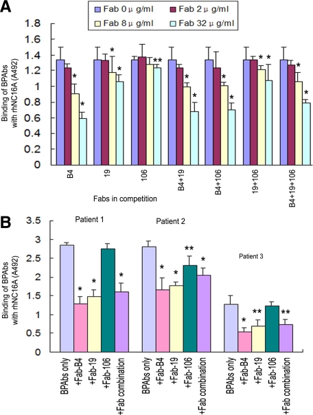
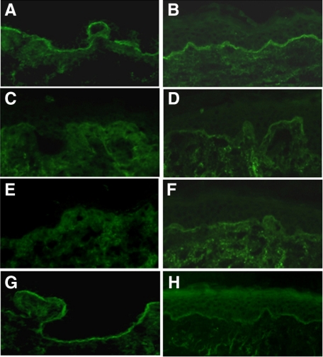
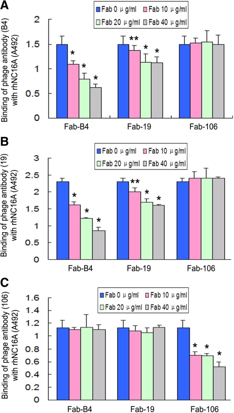
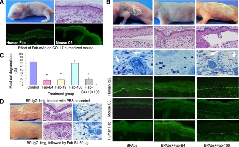
Activation of the complement cascade via the classical pathway is required for the development of tissue injury in many autoantibody-mediated diseases. It therefore makes sense to block the pathological action of autoantibodies by preventing complement activation through inhibition of autoantibody binding to the corresponding pathogenic autoantigen using targeted Fab antibody fragments. To achieve this, we use bullous pemphigoid (BP) as an example of a typical autoimmune disease. Recombinant Fabs against the non-collagenous 16th-A domain of type XVII collagen, the main pathogenic epitope for autoantibodies in BP, were generated from antibody repertoires of BP patients by phage display. Two Fabs, Fab-B4 and Fab-19, showed marked ability to inhibit the binding of BP autoantibodies and subsequent complement activation in vitro. In the in vivo experiments using type XVII collagen humanized BP model mice, these Fabs protected mice against BP autoantibody-induced blistering disease. Thus, the blocking of pathogenic epitopes using engineered Fabs appears to demonstrate efficacy and may lead to disease-specific treatments for antibody-mediated autoimmune diseases.


Figures

Figure 1
Figure 1
Isolation of specific binders against the NC16A domain of human type XVII collagen from phage antibody library. A: Randomly selected single colonies from the fourth panning round of the libraries show positive reaction with recombinant human NC16A (rhNC16A) but no reaction with recombinant mouse NC16A (rmNC16A), GST, or BSA in ELISA. B: Western blotting of soluble Fabs shows staining of only the rhNC16A domain and full-length human COL17, whereas rmNC16A and GST are negative. The representative results using Fab-B4 are shown. C: Epitope mapping of the generated Fabs with rhNC16A and its subdomains are shown. Fab-B4 binds to rhNC16A, NC16A2, and NC16A2.5 but does not react with NC16A1 and NC16A3, indicating that the binding epitope is located within an overlapping region within subdomain 2 and 2.5 (amino acids 514 to 520). The Fab-106 and Fab-19 epitopes are located in subdomain 1 (NC16A1, amino acids 490 to 506) and subdomain 2 (NC16A2, amino acids 507 to 520), respectively.
Figure 2
Figure 2
Production and characterization of Fabs. A: A purified soluble Fab in both reduced and nonreduced states is shown by Coomassie blue staining after SDS-PAGE. B: Immunogold labeling of normal human skin by Fabs shows 5-nm immunogold deposits restricted to immediately beneath hemidesmosomes, close to the keratinocyte plasma membrane (arrows, bar = 100 nm). The representative results using Fab-B4 are shown. (K: keratinocyte; D: dermis; KIF: keratin intermediate filaments; DEJ: dermal-epidermal junction). C: Immunofluorescence studies on normal human skin and skin sections from COL17 humanized mice show positive staining of the selected Fabs at the DEJ, and positive staining is also noted on the roof of NaCl split skin samples.
Figure 3
Figure 3
Inhibition ELISA assay. A: The effects of competition with Fab-B4, Fab-19, and various combinations inhibit the binding of autoantibodies (BPAbs, 8 μg/ml) from pooled sera of patients with bullous pemphigoid (BP) to rhNC16A in a dose-dependent manner (0 to 32 μg/ml), whereas Fab-106 inhibits BPAb binding only moderately. *P < 0.01, **P < 0.05, versus the original binding of BPAbs. B: Fabs (32 μg/ml) inhibit the binding of BPAbs from three BP patients. *P < 0.01, **P < 0.05, versus the original binding of BPAbs.
Figure 4
Figure 4
Inhibition immunofluorescence. A and B: Positive IgG staining of the NC16A affinity purified BPAbs (10 μg/ml) at the DEJ in human skin. C and E: IgG BPAbs staining is blocked by coincubation with either Fab-B4 or Fab-19 at a concentration of 20 μg/ml. G: Fab-106 (20 μg/ml) fails to significantly inhibit the binding of BPAbs. When BPAbs are allowed to bind to skin sections first and Fabs are added 30 minutes later, the IF staining of BPAb binding is also markedly reduced by Fab-B4 (D) or Fab-19 (F) but not by Fab-106 (H).
Figure 5
Figure 5
BPAb-induced complement activation and the inhibitory effects of Fabs. NC16A affinity purified BPAbs (10 μg/ml) induced activation of human C1q and C3 is shown at the DEJ in cryosections of human skin (A and B). When Fabs are coadministered with BPAbs at the same concentration, Fab-B4 completely blocks C1q and C3 activation (C and D), whereas Fab-19 effectively blocks the activation of C1q (E) and markedly reduces the activation of C3 (F). Fab-106 shows no inhibition of either C1q or C3 activation (G and H).
Figure 6
Figure 6
Inhibition ELISA for the three recombinant antibody clones using Fabs and phage antibodies (Phabs). Fab-B4 and Fab-19 inhibited the binding of both Phab-B4 and Phab-19 (A and B), whereas Fab-106 inhibited Phab-106 only (C). This indicates that the antibody clones B4 and 19 are mutually cross-inhibiting. *P < 0.01, **P < 0.05, versus the original binding of respective Phabs.
Figure 7
Figure 7
Therapeutic effects of Fabs on BP model mice. A: Results of the injection of Fabs into neonatal COL17 humanized mice show that this treatment alone does not cause BP disease or other detectable adverse effects. Histological examination (right, upper panel) supports this result. Indirect immunofluorescence show DEJ staining for the recombinant Fabs (left of lower panel) but no staining for mouse C3 (right, lower panel). B: Mice injected with NC16A affinity purified BPAbs develop the clinical and histological skin detachment associated with MC degranulation (white arrows) and the deposition of human IgG and mouse C3 at the DEJ. In contrast, mice injected with BPAbs and Fab-B4 fail to show these clinical and histological characteristics, and the intensity of IgG deposition at the DEJ is markedly reduced. The staining of mouse C3 is absent, whereas recombinant Fab fragment staining is weak but detectable. Fab-106 fails to show any beneficial therapeutic effect in the animal model. C: Percentage of dermal MC degranulation is assessed in BP model mice and in those treated with 30 μg/g body weight of Fabs. It is significantly reduced in the mice treated with Fab-B4, Fab-19, and the Fab combination. *P < 0.01 versus control group (BP model mice treated with PBS). D: BP model mice were produced by injection of BP-IgG (total IgG fraction prepared from BP patients) and were treated with Fab-B4 24 hours later. BP-like clinical and histological characteristics fail to develop in most (four of five) of Fab-B4 treated mice (lower panel).

Similar articles

Cited by

References

    1. Jacobson DL, Gange SJ, Rose NR, Graham NM. Epidemiology and estimated population burden of selected autoimmune diseases in the United States. Clin Immunol Immunopathol. 1997;84:223–243. - PubMed
    1. Holers VM. The complement system as a therapeutic target in autoimmunity. Clin Immunol. 2003;107:140–151. - PubMed
    1. Nishie W, Sawamura D, Goto M, Ito K, Shibaki A, McMillan JR, Sakai K, Nakamura H, Olasz E, Yancey KB, Akiyama M, Shimizu H. Humanization of autoantigen. Nat Med. 2007;13:378–383. - PubMed
    1. Liu Z, Giudice GJ, Swartz SJ, Fairley JA, Till GO, Troy JL, Diaz LA. The role of complement in experimental bullous pemphigoid. J Clin Invest. 1995;95:1539–1544. - PMC - PubMed
    1. Mewar D, Wilson AG. Autoantibodies in rheumatoid arthritis: a review. Biomed Pharmacother. 2006;60:648–655. - PubMed

Publication types

MeSH terms