. 2010 Mar;103(3):1283-94.
doi: 10.1152/jn.00704.2009. Epub 2009 Dec 30.

Long-lasting synaptic potentiation induced by depolarization under conditions that eliminate detectable Ca2+ signals

Affiliations

Long-lasting synaptic potentiation induced by depolarization under conditions that eliminate detectable Ca2+ signals

Fredy D Reyes et al. J Neurophysiol. 2010 Mar.

Abstract

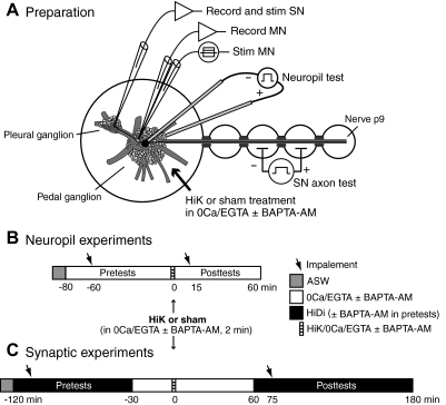
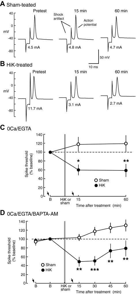
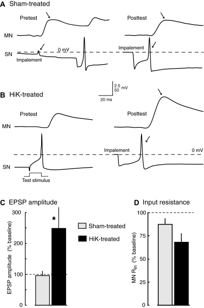
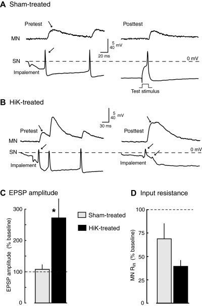
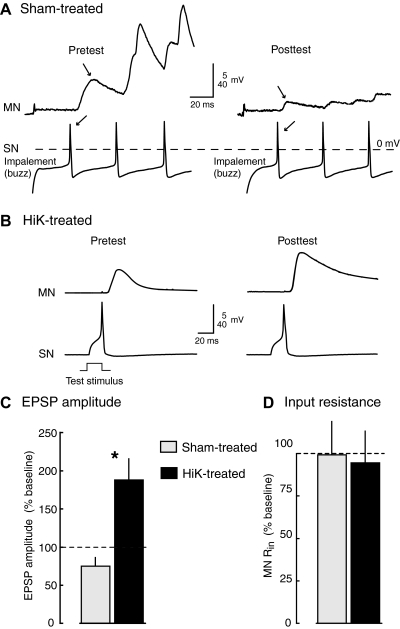
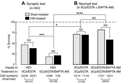
Activity-dependent alterations of synaptic transmission important for learning and memory are often induced by Ca(2+) signals generated by depolarization. While it is widely assumed that Ca(2+) is the essential transducer of depolarization into cellular plasticity, little effort has been made to test whether Ca(2+)-independent responses to depolarization might also induce memory-like alterations. It was recently discovered that peripheral axons of nociceptive sensory neurons in Aplysia display long-lasting hyperexcitability triggered by conditioning depolarization in the absence of Ca(2+) entry (using nominally Ca(2+)-free solutions containing EGTA, "0Ca/EGTA") or the absence of detectable Ca(2+) transients (adding BAPTA-AM, "0Ca/EGTA/BAPTA-AM"). The current study reports that depolarization of central ganglia to approximately 0 mV for 2 min in these same solutions induced hyperexcitability lasting >1 h in sensory neuron processes near their synapses onto motor neurons. Furthermore, conditioning depolarization in these solutions produced a 2.5-fold increase in excitatory postsynaptic potential (EPSP) amplitude 1-3 h afterward despite a drop in motor neuron input resistance. Depolarization in 0 Ca/EGTA produced long-term potentiation (LTP) of the EPSP lasting > or = 1 days without changing postsynaptic input resistance. When re-exposed to extracellular Ca(2+) during synaptic tests, prior exposure to 0Ca/EGTA or to 0Ca/EGTA/BAPTA-AM decreased sensory neuron survival. However, differential effects on neuronal health are unlikely to explain the observed potentiation because conditioning depolarization in these solutions did not alter survival rates. These findings suggest that unrecognized Ca(2+)-independent signals can transduce depolarization into long-lasting synaptic potentiation, perhaps contributing to persistent synaptic alterations following large, sustained depolarizations that occur during learning, neural injury, or seizures.


Figures

Fig. 1.
Fig. 1.
Preparation and experimental sequences. A: nerve-ganglia preparation consisting of excised pedal and pleural ganglia with the attached tail nerve (p9). Extracellular test stimuli were applied to the nerve to identify tail sensory neurons and then across the pedal ganglion (“neuropil test”) to determine the thresholds of their central processes in the vicinity of sensorimotor synapses (monitored by intracellular recording of evoked spikes conducted to the sensory neuron soma). Monosynaptic excitatory postsynaptic potentials (EPSPs) were tested by stimulating a tail sensory neuron (SN) and recording its synaptic response in a tail motor neuron (MN) that was manually clamped to −70 mV. MN input resistance (Rin) was tested by intracellular stimulation through a separate stimulating electrode. Neuropil test stimulation and MN recordings were performed in separate experiments. B: sequence of solution changes and microelectrode impalements into sensory neurons in experiments examining excitability of processes in the neuropil. The times indicated are relative to the offset of the 2-min high K+ (HiK) treatment. C: solution changes and SN and MN impalements in experiments examining synaptic potentiation. When present, BAPTA-AM was applied before and during the pretests, and was also included in the 0Ca/EGTA and high divalent cation (HiDi) solutions. In long-term experiments, the sequence was the same except that the posttests in HiDi were conducted 18–24 h after treatment.
Fig. 2.
Fig. 2.
Hyperexcitability of sensory neuron processes near central synaptic terminals induced by 2-min depolarization develops and is maintained in 0Ca/EGTA and 0Ca/EGTA/BAPTA-AM solutions. A and B: examples of sensory neuron action potentials evoked by threshold stimuli (stimulus current is indicated below each spike) before and after sham (A) and HiK (B) treatment. C: short- and intermediate-term hyperexcitability (STH/ITH) of sensory neuron processes in the pedal ganglion induced by 2-min treatment with HiK/0Ca/EGTA and maintained for ≥60 min in 0Ca/EGTA solution. Means of the median spike thresholds per preparation are normalized to the baseline (B) trial during the pretest phase (sham, n = 4 preparations with 10 sensory neurons; HiK, n = 4 preparations with 9 sensory neurons). Arrows indicate approximate times of microelectrode impalement. D: STH/ITH of sensory neuron processes in the pedal ganglion induced by treatment with HiK/0Ca/EGTA/BAPTA-AM and maintained in 0Ca/EGTA/BAPTA-AM solution (sham, n = 7 preparations with 11 sensory neurons; HiK, n = 5 preparations with 11 sensory neurons). For all excitability (and synaptic) data presented in this paper, each preparation contributed a single data point for each test (the median value from 1 to 4 sensory neurons tested per preparation) for statistical analyses. Differences between sham and HiK treatment outcomes were assessed with 2-way, repeated-measures ANOVA followed by Bonferroni posttests. *, P < 0.05; **, P < 0.01.
Fig. 3.
Fig. 3.
Potentiation of monosynaptic connections between tail sensory neurons and tail motor neurons is induced by 2-min conditioning depolarization under conditions that eliminate the driving force for Ca2+ entry into cells. A and B: examples of EPSPs evoked by the 1st action potential in sensory neurons stimulated in HiDi solution before (pretest) and ∼90 min after (posttest) treatment in sham (A) or HiK (B) solutions containing 0Ca/EGTA. In A, the 1st action potentials (←) and resulting EPSPs (→) are produced by impalement of the sensory neuron. No action potential occurred during impalement of the sensory neuron shown in B during the pretest, so the 1st action potential occurred when 20-ms pulses were delivered to the soma to determine soma spike threshold. C and D: significant potentiation of EPSP amplitude by HiK/0Ca/EGTA treatment. Graphs show means ± SE of the median test responses per preparation in each condition normalized to the 1st EPSP evoked by each sensory neuron during the pretest (sham, n = 9 preparations with 24 sensory neurons and 11 motor neurons; HiK, n = 7 preparations with 19 sensory neurons and 10 motor neurons). *, P < 0.05; **, P < 0.01, 2-tailed, unpaired t-test comparing the changes in responses (from pre- to posttest) between sham- and HiK-treated preparations.
Fig. 4.
Fig. 4.
Potentiation of monosynaptic connections between tail sensory neurons and tail motor neurons is induced by 2-min conditioning depolarization under conditions likely to eliminate Ca2+ transients during depolarization. A and B: examples of EPSPs evoked in HiDi solution before and ∼90 min after treatment in sham (A) or HiK (B) solutions containing 0Ca/EGTA/BAPTA-AM. In most cases, the 1st EPSP (→) was caused by sensory neuron spikes generated during impalement (←). EPSPs tested in HiDi solution before and 60–90 min after HiK/0Ca/EGTA/BAPTA-AM treatment showed significant potentiation compared with those tested after 0Ca/EGTA/BAPTA-AM treatment (C), while motor neuron input resistance tended to decrease (D; sham, n = 4 preparations with 13 sensory neurons and 6 motor neurons; HiK, n = 4 preparations with 7 sensory neurons and 4 motor neurons). *, P < 0.05, 1-tailed, unpaired t-test comparing the changes in responses (from pre- to posttest) between sham- and HiK-treated preparations. Graphs show means ± SE of the median test responses per preparation. One-tailed tests were utilized because the direction of the effect was predicted by the experiments summarized in Fig. 3 (see text).
Fig. 5.
Fig. 5.
Long-term synaptic potentiation is induced by 2-min conditioning depolarization under conditions that eliminate the driving force for Ca2+ entry. A and B: examples of EPSPs evoked in HiDi solution before and ∼20 h after treatment in sham (A) or HiK (B) solutions containing 0Ca/EGTA. The 1st sensory neuron spike (←) in each example in A was evoked during impalement facilitated by “buzzing” the microelectrode (oscillating the capacitance compensation circuit), which caused the artifacts at the beginning of each record. EPSPs (C) tested in HiDi solution before and 18–24 h after HiK/0Ca/EGTA treatment showed significant potentiation compared with those tested after sham treatment. Motor neuron Rin (D) was not altered in HiK- or sham-treated preparations in the long-term tests (sham, n = 3 preparations with 9 sensory neurons and 5 motor neurons; HiK, n = 4 preparations with 7 sensory neurons and 6 motor neurons). Graphs show means ± SE of the median test responses per preparation. *, P < 0.05, 2-tailed, unpaired t-test comparing the changes in responses overnight between sham- and HiK-treated preparations.
Fig. 6.
Fig. 6.
Conditions that reduce Ca2+ signaling increase sensory neuron mortality after re-exposure to high levels of extracellular Ca2+. A: survival rates of sensory neurons given synaptic tests in HiDi solution before and after exposure to low-Ca2+ solutions are sensitive to the degree of Ca2+ chelation but not to HiK treatment. Differences in the survival of HiK- and sham-treated sensory neurons were not sigifnicantly different (NS) in either condition, but the combined samples (HiK plus sham) of sensory neurons exposed to 0Ca/EGTA showed greater survival through the posttests than those exposed to 0Ca/EGTA/BAPTA-AM (43 of 101 vs. 19 of 100 cells, respectively, P < 0.001, ***, Fisher's exact test). B: survival rates of sensory neurons exposed to 0Ca/EGTA or 0Ca/EGTA/BAPTA-AM but not re-exposed to high levels of extracellular Ca2+ (during excitability tests in the neuropil, see Fig. 2). These were higher than those re-exposed to high Ca2+ levels after 0Ca/EGTA (B vs. A, left; 21 of 30 vs. 43 of 101 cells, respectively, P < 0.05, *) or 0Ca/EGTA/BAPTA-AM (B vs. A, right; 29 of 42 vs. 19 of 100 cells, respectively, P < 0.0001, ****). No significant differences were found in the survival rates of HiK- vs. sham-treated sensory neurons in 0Ca/EGTA or in 0Ca/EGTA/BAPTA-AM. These data were obtained from the experiments summarized in Figs. 2–4.

Similar articles

Cited by

References

    1. Abrams TW, Karl KA, Kandel ER. Biochemical studies of stimulus convergence during classical conditioning in Aplysia: dual regulation of adenylate cyclase by Ca2+/calmodulin and transmitter. J Neurosci 11: 2655–2665, 1991 - PMC - PubMed
    1. Antonov I, Antonova I, Kandel ER, Hawkins RD. The contribution of activity-dependent synaptic plasticity to classical conditioning in Aplysia. J Neurosci 21: 6413–6422, 2001 - PMC - PubMed
    1. Baba A, Yasui T, Fujisawa S, Yamada RX, Yamada MK, Nishiyama N, Matsuki N, Ikegaya Y. Activity-evoked capacitative Ca2+ entry: implications in synaptic plasticity. J Neurosci 23: 7737–7741, 2003 - PMC - PubMed
    1. Bailey CH, Giustetto M, Zhu H, Chen M, Kandel ER. A novel function for serotonin-mediated short-term facilitation in Aplysia: conversion of a transient, cell-wide homosynaptic hebbian plasticity into a persistent, protein synthesis-independent synapse-specific enhancement. Proc Natl Acad Sci USA 97: 11581–11586, 2000 - PMC - PubMed
    1. Ben-Chaim Y, Chanda B, Dascal N, Bezanilla F, Parnas I, Parnas H. Movement of “gating charge” is coupled to ligand binding in a G-protein-coupled receptor. Nature 444: 106–109, 2006 - PubMed

Publication types

MeSH terms

Substances

LinkOut - more resources