. 2010 Mar;30(3):498-508.
doi: 10.1161/ATVBAHA.109.200154. Epub 2009 Dec 30.

Diabetes mellitus induces bone marrow microangiopathy

Affiliations

Diabetes mellitus induces bone marrow microangiopathy

Atsuhiko Oikawa et al. Arterioscler Thromb Vasc Biol. 2010 Mar.

Abstract

Objective: The impact of diabetes on the bone marrow (BM) microenvironment was not adequately explored. We investigated whether diabetes induces microvascular remodeling with negative consequence for BM homeostasis.

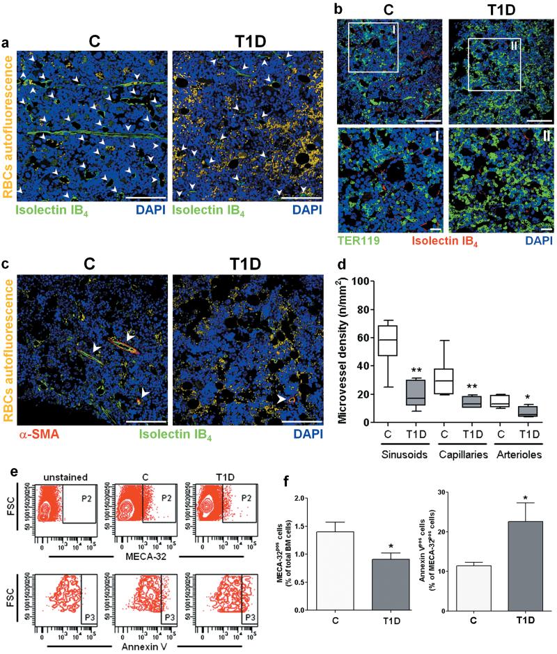
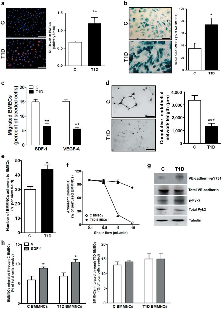
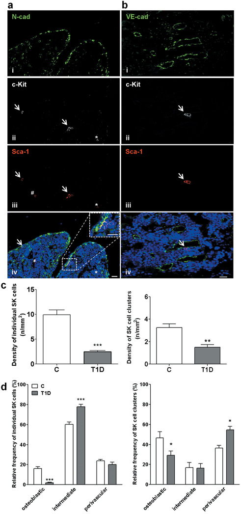
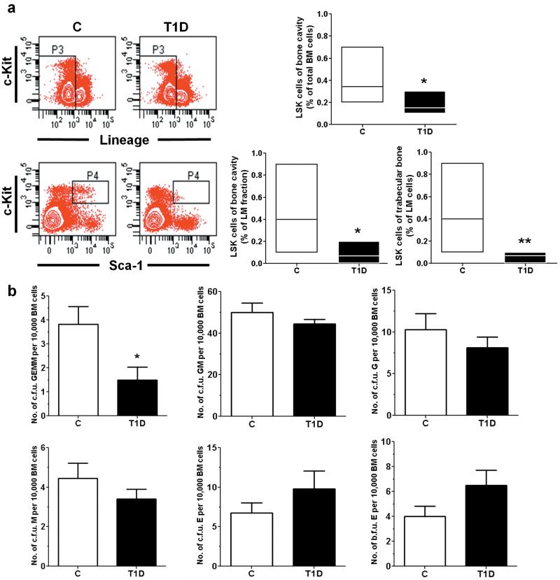
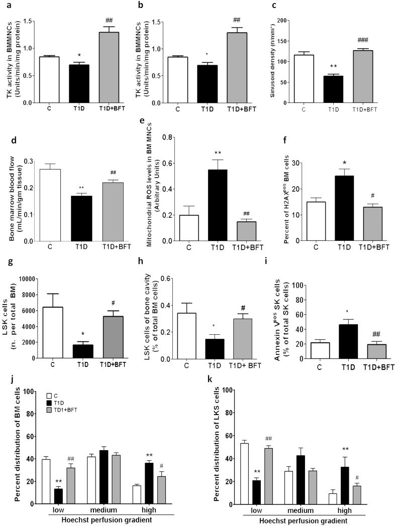
Methods and results: We found profound structural alterations in BM from mice with type 1 diabetes with depletion of the hematopoietic component and fatty degeneration. Blood flow (fluorescent microspheres) and microvascular density (immunohistochemistry) were remarkably reduced. Flow cytometry verified the depletion of MECA-32(+) endothelial cells. Cultured endothelial cells from BM of diabetic mice showed higher levels of oxidative stress, increased activity of the senescence marker beta-galactosidase, reduced migratory and network-formation capacities, and increased permeability and adhesiveness to BM mononuclear cells. Flow cytometry analysis of lineage(-) c-Kit(+) Sca-1(+) cell distribution along an in vivo Hoechst-33342 dye perfusion gradient documented that diabetes depletes lineage(-) c-Kit(+) Sca-1(+) cells predominantly in the low-perfused part of the marrow. Cell depletion was associated to increased oxidative stress, DNA damage, and activation of apoptosis. Boosting the antioxidative pentose phosphate pathway by benfotiamine supplementation prevented microangiopathy, hypoperfusion, and lineage(-) c-Kit(+) Sca-1(+) cell depletion.

Conclusions: We provide novel evidence for the presence of microangiopathy impinging on the integrity of diabetic BM. These discoveries offer the framework for mechanistic solutions of BM dysfunction in diabetes.


Figures

Figure 1
Figure 1. BM remodelling in T1D mice
(a) Representative images of H&E staining of femurs from C and T1D mice (scale bars: 500 μm). High magnifications of epiphysis and metaphysis show decreased cell density and empty spaces corresponding to fat accumulation in the marrow of the T1D mouse (scale bars: 100 μm). Box and whiskers graphs show min to max values of marrow volume (b), marrow cellular density (c), relative abundance of fat (d) and bone thickness (e). n=7 mice per group. *P<0.05 and **P<0.01 vs. C.
Figure 2
Figure 2. Microangiopathy in BM of T1D mice
Reduced vascular density and erythrocyte extravasation in T1D BM (a-d). Arrowheads point vascular structures. Scale bars: 100μm and 20μm (I and II). BMEC depletion and increased BMEC apoptosis in diabetes (e,f). n=8 mice per group. *P<0.05,**P<0.01 vs. C.
Figure 3
Figure 3. T1D-induced phenotypic alterations of BMECs
Microphotographs (scale bars: 100μm) and bar-graph illustrating ROS levels (a) and β-Gal activity (b) in BMECs. (c) Migration of BMECs toward SDF-1 and VEGF-A. (d) Endothelial network formation by BMECs plated on matrigel (Scale bars: 500μm). Adhesion of BMMNCs to non diabetic (C) BMECs or T1D BMECs under static conditions (e) and under the influence of shear flow (f). Western blot analysis of VE-cadherin-pY731 and Pyk2-pY402 (g). Trans-endothelial migration of BMMNCs towards SDF-1 (100 ng/mL) or vehicle (V) using BMECs isolated from C (h, left panel) or T1D mice (h, right panel) seeded on transwell inserts. For each assay, three separate experiments in triplicates were averaged. *P<0.05, **P<0.01 and ***P<0.001 vs. C.
Figure 4
Figure 4. T1D reduces the abundance of SK cells
Microphotographs (a,b) and graphs (c,d) showing SK cells of the osteoblastic (N-cad) and vascular niche (VE-cad). An individual cell (*) and clusters of cells (arrows) expressing c-Kit (ii) and Sca-1 (iii). Double-positive cells (purple fluorescence, iv). One cell expresses Sca-1 only (#). Scale bars: 20 μm. n=7 mice per group. *P<0.05,**P<0.01 and ***P<0.001 vs. C.
Figure 5
Figure 5. T1D depletes BM LSK cells
(a) Flow cytometry analysis of PIneg lineageneg c-Kitpos Sca-1pos cells. n=7 mice per group. (b) Colony forming unit (c.f.u.) assay of marrow cells harvested from trabecular bone. n=5 mice per group. *P<0.05, **P<0.01 vs. C.
Figure 6
Figure 6. Depletion of LSK cells follows perfusion gradient in diabetic BM
(a) Representative plots of Hoe uptake by BM cells and percent distribution of cells across the perfusion gradient. Abundance of LSK cells (b) and MECA32pos ECs (c) in each level of perfusion gradient. n=7 mice per group. *P<0.05, **P<0.01 vs. C.
Figure 7
Figure 7. Diabetes activates oxidative stress
(a) Intracellular ROS assessed by CM-H2DCFDA; *P<0.05,***P<0.001 vs. ROSlow, #P<0.05,##P<0.01 vs. C (b) Mitochondrial ROS assessed by MitoTracker Red CM-H2XROS. (c) Levels of p-H2AX (i: controls; ii: T1D. Scale bars=50μm). (d) Annexin Vpos SK cells. n=7 mice per group. *P<0.05,**P<0.01 and ***P<0.001 vs. C.
Figure 8
Figure 8. BFT prevents microangiopathy
Effect of BFT on transketolase (a) and G6PDH activity (b), sinusoid density (c), blood flow (d), ROS (e), and p-H2AX in BMMNCs (f). BFT prevents diabetes-induced depletion of LSK cells, assessed as absolute number (g) or percent of total BM cells (h), and reduces apoptosis (i). Bar graphs represent the percent of LSK cells in total BM cells (j) or LM (k) across the Hoe perfusion gradient. n=7 mice per group. *P<0.05,**P<0.01,***P<0.001 vs. C; #P<0.05 ##P<0.01, ###P<0.001 vs. vehicle.

Similar articles

Cited by

References

    1. Asahara T, Kawamoto A. Endothelial progenitor cells for postnatal vasculogenesis. Am J Physiol Cell Physiol. 2004;287:C572–579. - PubMed
    1. Dimmeler S. ATVB in focus: novel mediators and mechanisms in angiogenesis and vasculogenesis. Arterioscler Thromb Vasc Biol. 2005;25:2245. - PubMed
    1. Fischer C, Schneider M, Carmeliet P. Principles and therapeutic implications of angiogenesis, vasculogenesis and arteriogenesis. Handb Exp Pharmacol. 2006:157–212. - PubMed
    1. Jin DK, Shido K, Kopp HG, Petit I, Shmelkov SV, Young LM, Hooper AT, Amano H, Avecilla ST, Heissig B, Hattori K, Zhang F, Hicklin DJ, Wu Y, Zhu Z, Dunn A, Salari H, Werb Z, Hackett NR, Crystal RG, Lyden D, Rafii S. Cytokine-mediated deployment of SDF-1 induces revascularization through recruitment of CXCR4+ hemangiocytes. Nat Med. 2006;12:557–567. - PMC - PubMed
    1. Calvi LM, Adams GB, Weibrecht KW, Weber JM, Olson DP, Knight MC, Martin RP, Schipani E, Divieti P, Bringhurst FR, Milner LA, Kronenberg HM, Scadden DT. Osteoblastic cells regulate the haematopoietic stem cell niche. Nature. 2003;425:841–846. - PubMed

Publication types

MeSH terms