. 2010 Feb;23(2):377-86.

Fibroblasts from Werner syndrome patients: cancer cells derived by experimental introduction of oncogenes maintain malignant properties despite entering crisis

Affiliations

Fibroblasts from Werner syndrome patients: cancer cells derived by experimental introduction of oncogenes maintain malignant properties despite entering crisis

Furong Yuan et al. Oncol Rep. 2010 Feb.

Abstract

Werner syndrome (WS) results from defects in the gene encoding WRN RecQ helicase. WS fibroblasts undergo premature senescence in culture. Because cellular senescence is a tumor suppressor mechanism, we examined whether WS fibroblasts exhibited reduced tumorigenicity, in comparison to control cells, in a model of experimental conversion of normal human cells to cancer cells. The combination of oncogenic Ras (Ha-Ras(V12G)) and SV40 large T antigen (SV40 LT) causes human cells to acquire neoplastic properties in the absence of telomerase. We found that WS cells could also be converted to a tumorigenic state by these oncogenes, as evidenced by invasion and metastasis of cells implanted in immunodeficient mice. Ras/SV40 LT-expressing cells retained invasiveness and malignant properties even when cells reached crisis in tumors in vivo. High levels of gelatinase were found by an in situ assay in Ras/SV40 LT-expressing cells undergoing crisis. We conclude that, despite evidence of accelerated senescence in WS cells, there is no evidence that the absence of active WRN acts as a barrier to neoplastic transformation. Moreover, we find that tumorigenic human cells retain malignant properties of the cells as they approach and reach crisis.


Conflict of interest statement

Competing interests: The authors declare that they have no competing interests.

Figures

Figure 1
Figure 1. Malignant properties of WS fibroblasts expressing RasG12V and SV40 LT
Skin fibroblasts from four WS patients and three control subjects were transduced with retroviruses encoding RasG12V and SV40 LT and and were transplanted in the subrenal capsule space of immunodeficient mice. After 40 days mice were sacrificed and the gross appearance of tumors formed were photographed in situ using fluorescence under blue light. a-d, Werner syndrome donors (AG05229, AG04110, AG00780, AG06300); e-g, control donors (CRL-2707, CRL-2708, CRL-2714). a′-g′, Histological appearance of the tumors. Hematoxylin and eosin stain. Magnification a-g, x1; a′-g′, x200. The table gives the ages of the donors and tumor incidence. HCA2 are neonatal fibroblasts used in previous studies (19).
Figure 2
Figure 2. Loss of growth in serially transplanted tumors derived from WS and control fibroblasts
Ras/SV40 LT-transformed fibroblasts were transplanted in immunodeficient mice and allowed to grow for 40 days. Following sacrifice of the mice, the histology of the tumors was examined. Cells were also isolated from tumors and purified by flow cytometry, and then used in serial transplantation experiments. The serial transplantation histories of four reprentative tumors are shown in this figure. A hematoxylin/eosin-stained section of each tumor is shown. a and b show histories of tumors formed from control fibroblasts; c and d show histories of tumors formed from WS fibroblasts. In each case cells from a primary tumor were retransplanted into secondary host animals. Cells from secondary tumors were retransplanted into tertiary host animals. “X” indicates a failure of transplanted cells to form a detectable tumor. In some cases cells from primary or secondary tumors were infected with a retrovirus encoding hTERT, and were then retransplanted; tumors formed are indicated by 2°hT, etc., and were also serially transplanted. The dashed white line on tumor sections indicates the boundary between the kidney and the tumor tissue. In those cases where there is no dashed line the entire kidney was replaced by tumor. All tumor sections are shown at the same magnification, x1.
Figure 3
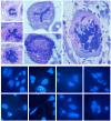
Figure 3. Crisis in tumors as evidenced by nuclear abnormalities
The top panel shows examples of abnormal cells in tumors derived from Ras/SV40 LT-expressing WS fibroblasts (hematoxylin- and eosin-stained sections). The top left example shows an anaphase bridge and other examples show cells with apparently abnormal numbers of spindles or other abnormal changes. The lower panel shows cells isolated from tumors and placed in culture (DAPI stain). The insert in the top left image shows an example of an anaphase bridge. Other cells show chromatin strings between separated nuclei, nuclear fragmentation, abnormal nuclear morphology or multiple nuclei. Magnifications: top panel, x500 and x1500; lower panel x500.
Figure 4
Figure 4. γ-H2AX in tumor cells in situ
a, b, c, Sections of tumors from Ras/SV40 LT-expressing WS fibroblasts were stained with an antibody against γ-H2AX. a′, b′, c′, DAPI stain. Magnification x500.
Figure 5
Figure 5. γ-H2AX in isolated tumor cells in culture
Cells were isolated from tumors from Ras/SV40 LT-expressing WS fibroblasts. a-e, stained with γ-H2AX antibody. a′-e′, DAPI stain. Insets show chromatin strings photographed with a higher exposure time.
Figure 6
Figure 6. Invasive characteristics of tumors formed from Ras/SV40 LT-expressing fibroblasts
Representative examples are shown to compare invasiveness of cells from WS and control donors. Hematoxylin and eosin stain. a, Primary tumor from LT/Ras-expressing WS fibroblasts. b,c Two secondary tumors derived by re-transplantation of cells from the tumor shown in a. d, Tertiary tumor derived by re-transplantation of the cells isolated from the tumor shown in b. e,f, Cells in crisis adjacent to kidney tubules (primary tumor from Ras/SV40 LT-expressing WS fibroblasts). g Primary tumor derived from LT/Ras-expressing control fibroblasts. h Secondary tumor derived by re-transplantation of cells isolated from the tumor shown in g. Magnification, a-d x50; e-f x200; g-h x50.
Figure 7
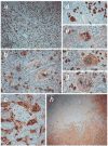
Figure 7. Tumor cells in crisis deep in the kidney parenchyma
Sections of tumors derived from Ras/SV40 LT-expressing fibroblasts were stained with anti-SV40 LT to enable identification of tumor cells within the kidney. a SV40 LT staining shows scattered crisis cells throughout the tumor. b, Crisis cells observed within the kidney adjacent to tubules and glomeruli. c, GFP staining demonstrates tumor cells surrounding a glomerulus. d-g, Examples of cells with abnormal nuclear morphology within the kidney adjacent to kidney tubules and glomeruli. h SV40 LT-staining in hTERT-transduced tumor cells retransplanted into immunodeficient mouse kidney. Magnification, a x50; b-g x200; h x25.
Figure 8
Figure 8. In situ gelatinase activity in fibroblasts before and after transduction with Ras/SV40 LT and in cells isolated from tumors
Cells were incubated with fluorescein-conjugated gelatin (DQ-gelatin) as described in Materials and Methods. All photographs were made with the same exposure time. a,a′, Nontransduced WS fibroblasts; b,b′, WS fibroblasts following transduction with Ras/SV40 LT; c,c′ Cells from tumor formed from Ras/SV40 LT-expressing WS fibroblasts; d,d′, Nontransduced control fibroblasts; e,e′, Control fibroblasts following transduction with Ras/SV40 LT; f,f′, Cells from tumor formed from Ras/SV40 LT-expressing control fibroblasts.

Similar articles

Cited by

References

    1. Martin GM. The Werner mutation: Does it lead to a “public” or “private” mechanism of aging? Mol Med. 1997;3:356–358. - PMC - PubMed
    1. Ostler EL, Wallis CV, Sheerin AN, Faragher RG. A model for the phenotypic presentation of Werner's syndrome. Exp Gerontol. 2002;37:285–92. - PubMed
    1. Goto M, Miller RW, Ishikawa Y, Sugano H. Excess of rare cancers in Werner syndrome (adult progeria) Cancer Epidemiol Biomarkers Prev. 1996;5:239–46. - PubMed
    1. Ishikawa Y, Miller RW, Machinami R, Sugano H, Goto M. Atypical osteosarcomas in Werner Syndrome (adult progeria) Jpn J Cancer Res. 2000;91:1345–9. - PMC - PubMed
    1. Brosh RM, Jr, Bohr VA. Human premature aging, DNA repair and RecQ helicases. Nucleic Acids Res. 2007;6:53–61. - PMC - PubMed

Publication types

MeSH terms

Substances